Achalasia: Symptoms, Causes, Types, Treatments
Understanding achalasia: Comprehensive guide to symptoms, diagnosis, and effective treatment options.

Understanding Achalasia: A Comprehensive Guide to Diagnosis and Treatment
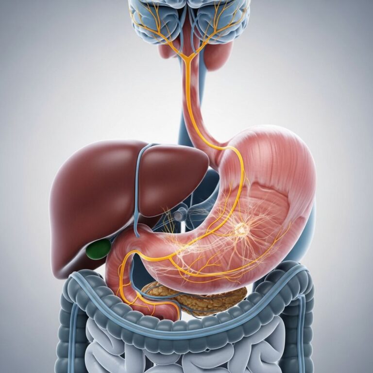
Achalasia, also known as cardiospasm, is a rare but serious condition that affects the esophagus, the muscular tube that carries food and liquids from your mouth to your stomach. In this disorder, the esophagus loses its ability to move food properly into the stomach due to dysfunction of the lower esophageal sphincter (LES), a ring of muscle that normally relaxes to allow food passage. When achalasia develops, this sphincter fails to relax appropriately, and the esophagus loses its normal peristaltic contractions—the coordinated muscle movements that push food downward. This combination creates a functional obstruction that prevents normal swallowing and digestion.
The condition is characterized by the buildup of food and liquid in the esophagus, which can lead to significant discomfort and nutritional challenges. Understanding achalasia is crucial because early diagnosis and appropriate treatment can prevent serious complications and dramatically improve quality of life. This guide explores the complete spectrum of achalasia, from its underlying causes and distinctive symptoms to the various diagnostic approaches and treatment modalities available.
What Is Achalasia?
Achalasia is an esophageal motility disorder that fundamentally alters how your esophagus functions. Unlike most swallowing disorders, achalasia specifically involves two major problems: the lower esophageal sphincter cannot relax properly, and the esophagus loses its ability to generate organized contractions that propel food downward. These dual dysfunctions create a mechanical obstruction at the junction between the esophagus and stomach.
The disorder is considered rare, affecting a small percentage of the population, though exact prevalence rates vary by geographic region. It can develop at any age but is most commonly diagnosed in adults. The condition is progressive, meaning that without treatment, symptoms typically worsen over time as the esophagus becomes progressively dilated from the accumulation of food and secretions.
Recognizing Achalasia Symptoms
The symptoms of achalasia typically develop gradually and can vary in severity from person to person. Understanding these signs is essential for early recognition and medical intervention. The most common symptoms include:
Dysphagia (Difficulty Swallowing) — This is the hallmark symptom of achalasia and typically affects both solids and liquids, though patients may notice more difficulty with solids initially. The sensation is often described as food becoming stuck in the chest or throat area. Patients may need to take multiple swallows, drink water to help push food down, or adopt specific postures to facilitate swallowing.
Regurgitation — Undigested food may regurgitate back up into the throat or mouth, particularly when lying down or shortly after eating. This regurgitation is often effortless and painless, distinguishing it from vomiting. Patients may experience sudden episodes of coughing or choking when food returns to the airway.
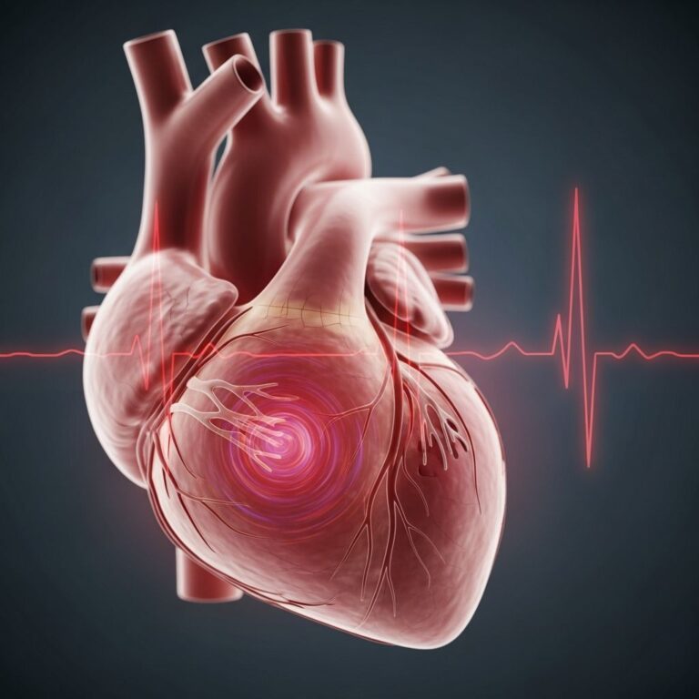
Chest Pain or Pressure — Some patients experience chest discomfort that can mimic cardiac problems. This pain may occur during or after meals and can range from mild pressure to significant discomfort. The similarity to cardiac pain often leads to cardiac workup before the true diagnosis is identified.
Heartburn and Reflux Symptoms — Despite the esophagus being blocked, patients may experience heartburn-like sensations due to fermentation of trapped food and bacterial overgrowth in the stagnant esophageal contents.
Weight Loss — Progressive weight loss often accompanies achalasia, sometimes occurring insidiously over months or years. This results from decreased food intake due to swallowing difficulties and potential malabsorption issues. Unintentional weight loss exceeding 10 percent of body weight may indicate serious disease progression.
Cough and Aspiration Risk — Regurgitated food can enter the airways, causing coughing, particularly at night or when lying down. This creates a risk for aspiration pneumonia, a serious complication where food or oral secretions enter the lungs.
Understanding the Types of Achalasia
Modern diagnostic techniques have identified distinct subtypes of achalasia based on specific pressure patterns and contraction characteristics observed during high-resolution manometry testing. These classifications help guide treatment decisions and predict treatment outcomes.
Type I Achalasia — Representing 20-40 percent of achalasia cases, Type I is characterized by failed peristalsis throughout the entire esophagus with minimal pressurization. This subtype typically presents with less severe symptoms and generally responds well to treatment, with good long-term outcomes.
Type II Achalasia — This is the most common form, accounting for 50-70 percent of cases. Type II features complete failed peristalsis combined with significant pressurization (greater than 30 mm Hg) in the esophageal body. Patients with Type II often experience more severe symptoms and may have greater esophageal dilation at diagnosis.
Type III Achalasia — The least common subtype, representing only 5 percent of cases, Type III is characterized by spastic contractions and abnormal pressure patterns. This type often presents with more atypical symptoms and may respond differently to standard treatments, sometimes requiring more aggressive intervention.
Diagnostic Approaches for Achalasia
Accurate diagnosis is fundamental to appropriate treatment planning. Healthcare providers employ several diagnostic tests to confirm achalasia and rule out other conditions that may mimic its symptoms.
High-Resolution Manometry (HRM) — This is the gold standard diagnostic test for achalasia. HRM measures the pressure patterns in your esophagus and sphincter with unprecedented precision. During this test, a thin pressure-sensitive tube is passed through your nose into your esophagus. The tube contains numerous sensors that measure pressure at different points as you swallow. The results create a detailed topographic map of esophageal pressures, allowing physicians to identify the specific dysfunction pattern and classify the achalasia subtype.
Barium X-Ray Studies — These imaging studies involve swallowing a barium contrast solution while X-rays capture images of the esophagus. In achalasia, characteristic findings include a dilated esophagus that gradually tapers to a narrow, tight junction at the lower esophageal sphincter, often described as a “bird’s beak” appearance. This test helps visualize the structural changes and degree of esophageal dilation.
Endoscopy — Upper endoscopy allows direct visualization of the esophageal lining and junction with the stomach. While endoscopy cannot definitively diagnose achalasia, it is valuable for excluding other conditions such as malignancy or strictures that might produce similar symptoms. During endoscopy, the gastroenterologist observes the appearance of the esophagus and lower esophageal sphincter, looking for secondary causes of obstruction.
Initial Clinical Assessment — Your healthcare provider will conduct a thorough physical examination and detailed symptom history, noting when symptoms began, their progression, associated factors, and impact on nutrition and daily activities.
Treatment Options for Achalasia
It is important to understand that achalasia is a chronic condition without a cure. All current treatment approaches are palliative, meaning they aim to relieve symptoms and improve esophageal function rather than eliminate the underlying disorder. Treatment goals focus on relaxing the lower esophageal sphincter, improving food passage, reducing symptoms, and preventing further esophageal complications.
Nonsurgical Treatment Approaches
Nonsurgical options provide initial symptom management and may be appropriate for patients who are not candidates for surgery or prefer conservative approaches initially.
Pharmacologic Therapy — Medications represent the least effective treatment option but may provide temporary symptom relief in some patients. Calcium channel blockers such as nifedipine (10-30 mg sublingual taken before meals) and nitrates like isosorbide dinitrate (5 mg sublingual before meals) are the most commonly used medications. These drugs work by reducing muscle tension in the lower esophageal sphincter. However, efficacy is limited, and many patients develop tolerance over time, requiring dose escalation or discontinuation.
Botulinum Toxin (Botox) Injections — Botox can be injected directly into the lower esophageal sphincter during endoscopy to temporarily paralyze the muscle. This typically provides symptom relief for 3-6 months, after which injections must be repeated. While Botox offers a minimally invasive option, repeat procedures may eventually cause antibody formation, reducing effectiveness.
Pneumatic Dilation — This endoscopic procedure involves advancing a balloon catheter across the lower esophageal sphincter and inflating it with controlled pressure to disrupt the muscle fibers. Pneumatic dilation is effective for short and long-term symptom relief in many patients. However, the procedure carries a 1.9 percent risk of esophageal perforation, a serious complication requiring surgical repair. Patients who undergo pneumatic dilation must be surgical candidates in case perforation occurs. Additionally, symptoms may recur over time, necessitating repeat procedures.
Surgical Treatment Options
Surgical approaches offer more definitive and longer-lasting relief compared to nonsurgical options and are considered among the most effective treatments available.
Laparoscopic Heller Myotomy (LHM) — This minimally invasive surgical procedure involves division of the circular muscle fibers of the lower esophageal sphincter without disrupting the mucosa (inner lining). The procedure is performed laparoscopically through several small incisions in the abdomen, offering faster recovery compared to open surgery. The surgeon carefully divides the muscle fibers on the anterior aspect of the esophagus, typically 6 centimeters above and 2-3 centimeters below the gastroesophageal junction. A partial fundoplication (wrapping of stomach tissue) is often added to prevent postoperative reflux. Laparoscopic Heller myotomy has shown excellent long-term success rates, with 60-94 percent of patients achieving good-to-excellent results that persist for many years.
POEM (Peroral Endoscopic Myotomy) — This newer minimally invasive technique represents an innovative approach to achalasia treatment. POEM is performed entirely endoscopically, without surgical incisions. During the procedure, the endoscope is guided into the esophagus, and a tunnel is created in the submucosa (layer beneath the inner lining). A specialized knife then divides the circular muscle fibers along this tunnel while preserving the mucosa. After completing the myotomy, endoscopic clips seal the mucosal entry site. POEM offers excellent short and long-term outcomes comparable to laparoscopic myotomy, with the advantages of no external incisions and potentially faster recovery.
Esophagectomy — In rare cases where symptoms are severe and other treatments have failed or are not suitable, esophagectomy (surgical removal of the esophagus) may be considered. This is a major procedure reserved for end-stage disease and is seldom necessary with modern treatment options.
Comparative Effectiveness of Treatment Modalities
| Treatment | Effectiveness | Duration | Invasiveness | Complications |
|---|---|---|---|---|
| Medications | Low | Variable/temporary | None | Minimal |
| Botox | Moderate | 3-6 months | Minimally invasive | Antibody resistance |
| Pneumatic Dilation | High | 1-5+ years | Minimally invasive | Perforation (1.9%) |
| Laparoscopic Myotomy | High | Long-term (years) | Minimally invasive | Reflux, bleeding |
| POEM | High | Long-term (years) | Minimally invasive | Reflux, mucosal injury |
Recovery and Follow-Up Care
Recovery experiences vary depending on the treatment modality selected. For minimally invasive procedures like pneumatic dilation, POEM, or laparoscopic myotomy, most patients experience relatively quick recovery compared to open surgery.
After endoscopic procedures such as POEM or pneumatic dilation, patients typically remain hospitalized overnight for observation. The morning after the procedure, a barium X-ray is performed to confirm proper esophageal function and ensure no leakage has occurred. Patients are started on a liquid diet and usually discharged later that day. Follow-up with your surgeon occurs 7-10 days after the procedure, with additional swallow studies performed around the three-month mark to verify continued proper esophageal emptying.
Long-term follow-up is essential because achalasia symptoms frequently recur after treatment. Your healthcare provider will monitor for symptom recurrence, assess esophageal function over time, and watch for potential complications such as reflux disease or esophageal dilation.
Potential Complications Without Treatment
Untreated achalasia can lead to serious complications that significantly impact health and quality of life. Progressive malnutrition from inadequate food intake can become life-threatening and affect life expectancy. Aspiration pneumonia may develop when regurgitated food enters the lungs. The esophagus can become severely dilated, a condition called megaesophagus. Additionally, there is a slightly elevated risk of esophageal cancer in patients with long-standing, untreated achalasia, though this remains relatively uncommon.
With appropriate treatment, however, patients can expect to maintain normal life expectancy and quality of life comparable to individuals without the disorder.
Frequently Asked Questions About Achalasia
Q: Can achalasia be cured?
A: No, achalasia cannot be cured with current medical knowledge. However, all available treatments are effective at managing symptoms and improving esophageal function. Treatment goals focus on symptom relief and preventing complications rather than eliminating the underlying condition.
Q: Which treatment is best for achalasia?
A: The best treatment depends on individual factors including symptom severity, age, overall health status, esophageal anatomy, and personal preferences. Both pneumatic dilation and laparoscopic myotomy have demonstrated equivalent long-term effectiveness. Your healthcare provider will discuss all options to determine the most appropriate choice for your specific situation.
Q: How is achalasia diagnosed?
A: High-resolution manometry is the gold standard diagnostic test. It measures pressure patterns in the esophagus and lower esophageal sphincter. Your doctor may also use barium X-rays and endoscopy to support the diagnosis and rule out other conditions.
Q: What are the risks of achalasia treatment?
A: Each treatment carries specific risks. Pneumatic dilation has a 1.9 percent risk of esophageal perforation. Surgical approaches may result in reflux disease or bleeding. Botox requires repeated procedures and may lose effectiveness over time. Discuss specific risks for your chosen treatment with your healthcare provider.
Q: Can achalasia symptoms recur after treatment?
A: Yes, symptoms often recur after treatment, particularly with pneumatic dilation and Botox injections. This is why long-term follow-up care is essential. Your healthcare provider will monitor your condition and discuss repeat treatment if symptoms return.
Q: How does POEM differ from laparoscopic myotomy?
A: POEM is performed entirely through the endoscope without external incisions, while laparoscopic myotomy requires small surgical incisions in the abdomen. Both procedures have excellent outcomes, but POEM may offer faster recovery and fewer external wounds. Your doctor can help determine which approach suits your situation best.
References
- ACG Clinical Guidelines: Diagnosis and Management of Achalasia — American College of Gastroenterology. 2022. https://pubmed.ncbi.nlm.nih.gov/32773454/
- Achalasia (Cardiospasm): Symptoms, Causes, Types, Treatments — Cleveland Clinic. 2025. https://my.clevelandclinic.org/health/diseases/17534-achalasia
- POEM: Treatment Procedure For Achalasia — Cleveland Clinic. 2025. https://my.clevelandclinic.org/departments/digestive/depts/poem
- Esophageal Motility Disorders and Achalasia Subtypes — National Center for Biotechnology Information. 2023. https://pmc.ncbi.nlm.nih.gov/articles/PMC9896940/
Similar Articles


Read full bio of Sneha Tete