Acrokeratoelastoidosis: Symptoms, Causes, And Treatment Guide
Rare genetic skin disorder causing keratotic papules on hands and feet: causes, diagnosis, and management options.

Author: Expert Dermatologist (Perplexity AI Synthesis)
Revised: January 2026
What is acrokeratoelastoidosis?
Acrokeratoelastoidosis (AKE), also known as acrokeratoelastoidosis of Costa, is a rare genetic dermatosis characterized by the development of small, firm, umbilicated, skin-coloured or yellowish keratotic papules predominantly along the borders of the hands and feet. First described by Brazilian dermatologist Oswaldo Costa in 1952, this condition falls under the spectrum of marginal papular acrokeratodermas (MPA), which feature keratotic papules on acral margins. It typically manifests during late childhood or puberty, progressing gradually before stabilizing, and is inherited in an autosomal dominant manner with variable penetrance.
The papules are usually asymptomatic, causing no pain, itching, or functional impairment, but they can be cosmetically distressing for affected individuals. Lesions are confined to acral areas, sparing the central palms and soles in classic presentations, though atypical involvement may occur. AKE is benign with an excellent prognosis, showing no association with malignancy or systemic disease, and spontaneous resolution is not reported.
Who gets acrokeratoelastoidosis?
Acrokeratoelastoidosis primarily affects individuals of all ages but commonly presents in the second or third decade of life, with onset during puberty or early adulthood in many cases. It exhibits a female predominance in some reported series, though this may reflect reporting bias rather than true epidemiology. Familial cases underscore its autosomal dominant inheritance, with pedigrees showing vertical transmission across generations.
Geographic clustering has been noted in certain populations, such as those of European descent or in regions like South America where it was first identified, but it occurs worldwide without ethnic predisposition. No environmental triggers like sun exposure are required, distinguishing it from actinic damage-related conditions. Patients often have a family history, with up to 40-60% of cases reported as hereditary in literature reviews.
What causes acrokeratoelastoidosis?
The precise etiology remains elusive, but AKE is widely regarded as a genetic disorder linked to abnormalities in elastic fiber metabolism and keratinization. Histopathological hallmarks suggest defective elastic fiber secretion by dermal fibroblasts rather than primary degradation, leading to elastorrhexis—fragmentation and reduction of elastic fibers in the dermis. Excessive filaggrin accumulation in the stratum granulosum may contribute to keratotic papule formation.
- Genetic factors: Autosomal dominant inheritance with incomplete penetrance; specific gene mutations (e.g., in elastin-related pathways) are hypothesized but not definitively identified.
- Pathophysiological mechanisms: Abnormal dermal collagen homogenization, hyperkeratosis, and hypergranulosis; repeated microtrauma may exacerbate acral predominance.
- Associations: Rare links to scleroderma-like connective tissue changes or hyperhidrosis, but no causal relationship established.
No infectious, autoimmune, or malignant drivers have been implicated, reinforcing its idiopathic genetic basis.
What are the clinical features of acrokeratoelastoidosis?
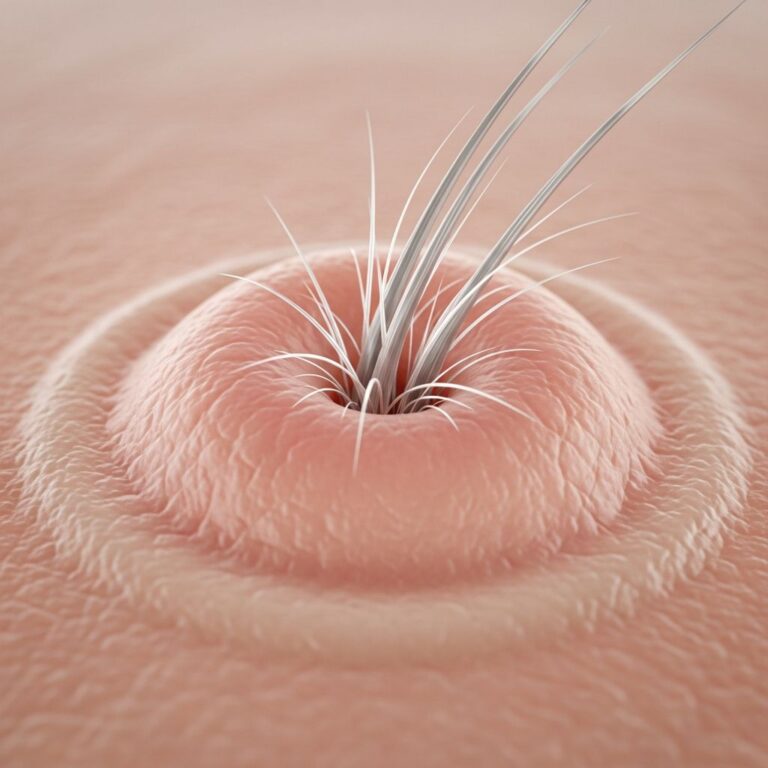
Classic lesions are 1-3 mm firm, warty papules with central umbilication, appearing yellowish, flesh-colored, or translucent, coalescing into plaques along the lateral margins of palms, fingers, soles, and toes. The dorsal hands and feet backs may also be involved, with structureless pale yellow patches interspersed.
Symptoms are minimal: dryness or mild scaling, but rarely pruritus or tenderness. Progression halts after initial development, remaining stable lifelong without scarring or atrophy. Atypical presentations include plaque forms or involvement of non-acral sites, as in rare adult-onset cases.
| Feature | Description | Frequency |
|---|---|---|
| Papules | Firm, umbilicated, 1-3mm on acral borders | Common (>90%) |
| Color | Skin-colored to yellowish | Common |
| Symptoms | Asymptomatic or dry | Most cases |
| Distribution | Hands/feet margins | Exclusive in classic form |
Pathology of acrokeratoelastoidosis
Histopathology confirms diagnosis, revealing compact hyperkeratosis, hypergranulosis, mild acanthosis, and superficial dermal changes. Key features include collagen homogenization, reduced/fragmented elastic fibers (elastorrhexis) on Verhoeff-Van Gieson or aldehyde fuchsin stains, without inflammation or vasculitis.
- Epidermis: Hyperkeratosis with occasional parakeratosis; dense filaggrin bands in granulosum.
- Dermis: Elastorrhexis, hyalinized collagen, sparse fibroblasts with dense granules.
These findings distinguish AKE from mimics like porokeratosis or viral warts.
Diagnosis of acrokeratoelastoidosis
Diagnosis is primarily clinical, based on age of onset (post-puberty), family history, acral distribution without photodamage, and biopsy confirmation. Dermoscopy may show umbilication and keratotic plugs.
Differential diagnoses include:
- Punctate palmoplantar keratoderma
- Porokeratosis of Mibelli
- Common warts
- Focal acral hyperkeratosis
- Digital papular eccrine ostial and dermal duct nevus
Treatment of acrokeratoelastoidosis
No cure exists due to its genetic nature; treatment is optional for cosmetic concerns as lesions are stable and asymptomatic. Options include:
| Category | Examples | Efficacy | Notes |
|---|---|---|---|
| Topical | Salicylic acid, urea, tretinoin, corticosteroids | Mild | Relieves dryness; recurrence common |
| Systemic | Acitretin, methotrexate, dapsone, prednisolone | Moderate (acitretin best) | Relapse on discontinuation; side effects |
| Physical | Er:YAG laser, cryotherapy | Variable | Limited benefit; no recurrence in some laser cases |
Watchful waiting is recommended for most, with counseling on benign prognosis.
Frequently asked questions about acrokeratoelastoidosis
Is acrokeratoelastoidosis contagious?
No, it is a genetic condition, not infectious.
Does acrokeratoelastoidosis go away?
No, lesions stabilize but persist lifelong without spontaneous resolution.
Can acrokeratoelastoidosis be cured?
No cure; management is symptomatic.
Is acrokeratoelastoidosis painful?
Rarely; mostly cosmetic issue.
What is the prognosis for acrokeratoelastoidosis?
Excellent; no systemic risks.
References
- Acrokeratoelastoidosis – PMC – NIH — National Library of Medicine. 2015. https://pmc.ncbi.nlm.nih.gov/articles/PMC4693374/
- Acrokeratoelastoidosis of Costa – Wikipedia — Wikipedia Contributors. 2023-10-01. https://en.wikipedia.org/wiki/Acrokeratoelastoidosis_of_Costa
- Acrokeratoelastoidosis of Costa: Clinical Image — Athenaeum Publishing. 2020. https://athenaeumpub.com/wp-content/uploads/Acrokeratoelastoidosis-of-Costa-Clinical-Image.pdf
- Acrokeratoelastoidosis – DermNet — DermNet NZ. 2024. https://dermnetnz.org/topics/acrokeratoelastoidosis
- Acrokeratoelastoidosis (Genetic Skin Lesions) — MD Searchlight. 2023. https://mdsearchlight.com/genetic-disorders/acrokeratoelastoidosis-genetic-skin-lesions/
- Plaques: A Rare Presentation of Acrokeratoelastoidosis — The Hospitalist. 2022. https://blogs.the-hospitalist.org/content/plaques-rare-presentation-acrokeratoelastoidosis
- Acrokeratoelastoidosis – StatPearls — NCBI Bookshelf. 2023-07-17. https://www.ncbi.nlm.nih.gov/books/NBK537067/
Similar Articles


Read full bio of medha deb