Adie Syndrome: Understanding Pupil Dysfunction
A comprehensive guide to recognizing and managing Adie syndrome symptoms

Adie syndrome, also referred to as Holmes-Adie syndrome or tonic pupil disorder, represents a distinctive neurological condition that disrupts the normal functioning of the eye’s pupil and certain reflexes throughout the body. This rare disorder affects the autonomic nervous system, which controls involuntary bodily functions including pupil constriction and dilation. Understanding this condition is essential for individuals who may experience unusual pupil behavior or vision changes, as early recognition can lead to appropriate management and symptom relief.
Understanding the Core Mechanism
The pupil, the dark circular opening in the center of the eye, plays a crucial role in regulating the amount of light that enters the eye. Under normal circumstances, the pupil automatically constricts in response to bright light and dilates in dim conditions. This automatic response occurs through the parasympathetic nervous system, specifically through nerve connections that pass through the ciliary ganglion, a small nerve cluster located behind the eye.
In Adie syndrome, the nerves responsible for controlling pupil constriction become damaged or inflamed, disrupting this normal reflex pathway. When these parasympathetic nerves malfunction, the affected pupil becomes abnormally dilated and fails to respond appropriately to light exposure. The condition typically affects one eye initially, though it may eventually progress to involve both eyes over time.
Recognizing the Primary Characteristics
The hallmark feature of Adie syndrome is the presence of one pupil that appears noticeably larger than the other, a condition known as anisocoria. This enlargement becomes particularly evident in normal room lighting. The affected pupil demonstrates several distinctive characteristics:
- Persistent dilation even when exposed to bright light
- Slow or minimal constriction in response to direct light stimulation
- More pronounced constriction when focusing on nearby objects compared to response to light
- A tendency to become slightly oval or irregular in shape rather than perfectly round
- Gradual reduction in size over months or years, though light responsiveness typically does not fully recover
This light-near dissociation—where the pupil responds better to accommodation (focusing on close objects) than to light—distinguishes Adie syndrome from other pupil-related conditions. The nerve damage also triggers the pupil to develop supersensitivity to acetylcholine, a neurotransmitter involved in muscle contraction, resulting in the slow and sustained constriction response characteristic of the tonic pupil.
Associated Symptoms and Visual Complaints
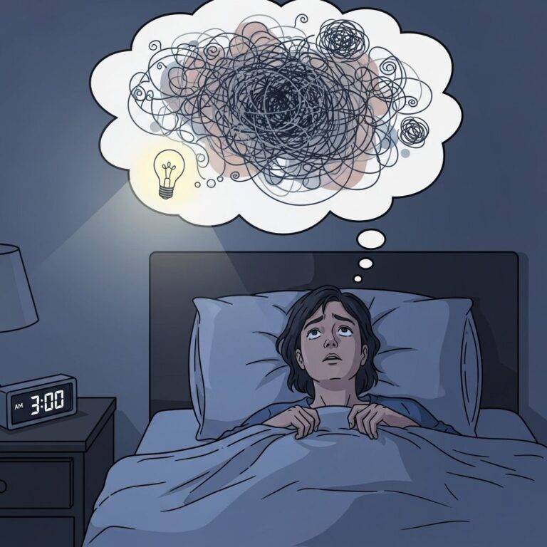
While the abnormal pupil represents the most visible sign of Adie syndrome, patients often experience a range of accompanying symptoms that affect daily functioning. Photophobia, or extreme sensitivity to bright light, stands as the most commonly reported complaint. Because the affected pupil cannot constrict normally, excessive light reaches the retina, causing discomfort and an instinctive desire to close the eyes or squint when exposed to sunlight or bright indoor lighting.
Vision-related symptoms frequently include:
- Blurred vision at various distances, particularly in the affected eye
- Difficulty focusing on objects at close range, especially when reading or performing detailed tasks
- Impaired depth perception, affecting the ability to judge distances accurately
- Glare and halos around light sources, particularly noticeable at night or in low-light conditions
- Reduced ability to adapt when transitioning between different lighting environments
The severity and combination of symptoms varies considerably among individuals. Some people experience minimal visual difficulties, while others find that photophobia and focusing problems substantially impact their quality of life. It is important to note that these symptoms typically originate from the affected eye, though bilateral involvement can develop over time.
Neurological Manifestations Beyond the Eye
Adie syndrome frequently extends beyond ocular symptoms to include disruptions in reflexes throughout the body. Deep tendon reflexes, the automatic muscle contractions that occur when a healthcare provider taps a tendon with a reflex hammer, may be absent or significantly diminished. The Achilles tendon reflex, tested by tapping the back of the ankle, represents the most commonly affected reflex, though the patellar reflex at the knee may also be impaired.
These reflex abnormalities result from damage to the dorsal root ganglia, nerve clusters where sensory nerve fibers originate along the spinal cord. The same inflammatory or degenerative process affecting the ciliary ganglion can compromise these other nerve structures, leading to the broader neurological presentation that defines Holmes-Adie syndrome specifically. When Adie syndrome occurs without reflex abnormalities, the condition may be referred to simply as Adie tonic pupil.
Investigating Potential Causes
In the majority of cases—approximately 80 percent—Adie syndrome develops without an identifiable cause, classified as idiopathic. However, researchers have identified several conditions and events that can trigger the development of a tonic pupil. These causative factors include:
| Potential Trigger | Mechanism of Action |
|---|---|
| Viral Infections | Inflammatory response damages nerve fibers |
| Traumatic Injury | Direct damage to autonomic nerve pathways |
| Surgical Procedures | Unintended nerve injury during eye or nearby surgery |
| Tumors | Mass effect or compression of nerve structures |
| Reduced Blood Flow (Ischemia) | Insufficient oxygen supply damages nerve tissue |
| Autoimmune Conditions | Immune system attacks nerve fibers and ganglia |
| Vasospasm from Migraines | Blood vessel constriction reduces oxygen availability |
Additionally, Adie tonic pupil can occur as a feature of other systemic conditions affecting the autonomic nervous system, including Ross syndrome (which combines tonic pupil with segmental anhidrosis, or reduced sweating) and Harlequin syndrome. Understanding the potential underlying cause informs both the diagnostic approach and long-term management strategy.
Diagnostic Procedures and Testing
Diagnosis of Adie syndrome begins with a comprehensive eye examination and detailed patient history. An ophthalmologist or optometrist will observe the pupil size in various lighting conditions, noting the characteristic enlargement and reduced light responsiveness. The examiner may also perform slit-lamp biomicroscopy, which uses a specialized microscope to examine the eye closely and may reveal sectoral iris sphincter paralysis, where portions of the iris muscle fail to contract normally, creating a vermiform or worm-like movement of the pupillary margin.
A critical diagnostic test involves assessing light-near dissociation by comparing the pupil’s response to bright light with its response to accommodation. The examiner shines a light in the eye and asks the patient to focus on a nearby object, noting whether the pupil constricts more effectively with near accommodation than with light exposure.
The pharmacological test using dilute pilocarpine drops represents one of the most specific diagnostic tools. Pilocarpine, which mimics acetylcholine, causes the tonic pupil of Adie syndrome to constrict at very low concentrations (0.05-0.1 percent) that would not affect a normal pupil. This supersensitivity to dilute pilocarpine, combined with failure to respond to stronger concentrations of other drugs, helps distinguish Adie syndrome from other causes of pupil dilation.
Testing deep tendon reflexes through physical examination confirms whether reflex loss accompanies the pupil abnormality. Some patients may undergo additional imaging or laboratory studies if the clinical presentation suggests an underlying systemic condition rather than idiopathic Adie syndrome.
Management and Treatment Strategies
Currently, no treatment reverses the underlying nerve damage in Adie syndrome; however, multiple approaches can effectively manage symptoms and improve visual comfort and function. The treatment plan should be individualized based on the specific symptoms each patient experiences.
For photophobia management, protective eyewear plays a central role. Tinted or dark glasses reduce light reaching the affected eye, significantly decreasing discomfort in bright environments. Specialized photochromic lenses, which darken automatically in response to light exposure, offer convenient protection in varying lighting conditions. Some individuals benefit from specific lens tints designed to reduce glare and enhance contrast.
Vision problems, particularly difficulty focusing at near distances, may be addressed through corrective glasses prescribed specifically for reading or close work. Bifocals or progressive lenses can help optimize vision across multiple distances. In some cases, lubricating eye drops help alleviate dry eye symptoms that may accompany the visual dysfunction.
Recent research has explored the use of pilocarpine drops as a therapeutic intervention. By applying dilute pilocarpine to the affected eye, the supersensitive tonic pupil may constrict further, reducing the amount of light entering the eye and potentially alleviating photophobia. However, the long-term efficacy and appropriate dosing regimens continue to be investigated.
Prognosis and Long-Term Outlook
The long-term prognosis for individuals with Adie syndrome is generally favorable from the perspective of vision loss or serious complications. The condition itself does not lead to blindness or progressive deterioration in visual acuity. However, several important trends characterize the disease course. Over months to years, the affected pupil tends to gradually decrease in size, though it rarely returns to normal dimensions. Light responsiveness generally does not improve substantially over time, despite changes in resting pupil size.
Approximately four percent of individuals per year experience progression of the condition to the second eye, with roughly 50 percent developing bilateral involvement within five to ten years. When both eyes are affected, the condition becomes more symmetrical, potentially reducing the noticeable asymmetry of pupil size.
The reflex abnormalities associated with Holmes-Adie syndrome appear to be nonprogressive in most cases. However, the presence of reflex loss may increase the likelihood of developing other autonomic nervous system conditions, warranting periodic medical evaluation.
When to Seek Professional Evaluation
Any individual noticing a persistent difference in pupil size between the two eyes, unexplained photophobia, or changes in focusing ability should consult with an eye care professional. While Adie syndrome itself is not vision-threatening, similar pupil abnormalities can indicate other neurological conditions requiring different management approaches. A thorough examination ensures accurate diagnosis and appropriate treatment planning.
Patients already diagnosed with Adie syndrome should maintain regular follow-up appointments to monitor for progression to the second eye and to reassess symptoms and treatment effectiveness. Any new neurological symptoms should be reported promptly to allow for comprehensive evaluation.
References
- Tonic Pupil (Adie Syndrome) — American Optometric Association. https://www.optometrists.org/general-practice-optometry/guide-to-eye-health/conditions-that-affect-the-pupil/tonic-pupil-adie-syndrome/
- Adie Pupil — EyeWiki. American Academy of Ophthalmology. https://eyewiki.org/Adie_Pupil
- Adie Syndrome (Tonic Pupil): Causes, Symptoms & Treatment — Cleveland Clinic. https://my.clevelandclinic.org/health/diseases/adie-syndrome-tonic-pupil
- Adie Syndrome – Symptoms, Causes, Treatment — National Organization for Rare Disorders (NORD). https://rarediseases.org/rare-diseases/adie-syndrome/
- Adie Syndrome: Treatment, Symptoms, and Causes — Medical News Today. https://www.medicalnewstoday.com/articles/adie-syndrome
Similar Articles


Read full bio of Sneha Tete