Adnexal Tumours: Types, Diagnosis, and Management
Comprehensive guide to cutaneous adnexal tumours: benign and malignant skin conditions.

Adnexal Tumours: Comprehensive Overview
Cutaneous adnexal tumours are a diverse group of benign and malignant neoplasms that originate from the normal structures found within the skin. These tumours arise from three primary sources: hair follicles, sebaceous glands, and eccrine and apocrine sweat glands. Understanding the nature, classification, and management of these conditions is essential for dermatologists and healthcare professionals involved in skin cancer diagnosis and treatment.
What Are Adnexal Tumours?
Adnexal tumours represent a large and heterogeneous group of skin lesions that exhibit morphological differentiation toward one or more types of adnexal epithelium present in normal skin. These structures include the pilosebaceous unit (hair follicles and sebaceous glands) as well as eccrine and apocrine sweat glands. The vast majority of adnexal tumours encountered in clinical practice are benign, though malignant variants exist and require careful evaluation.
Most skin adnexal tumours are relatively uncommonly encountered in routine dermatological practice. This relative rarity can present diagnostic challenges, as pathologists may have limited exposure to frequently encountered tumours within this category. The diagnosis of adnexal tumours relies predominantly on histological evaluation rather than clinical presentation alone, making accurate tissue sampling and pathological interpretation critical.
Classification and Origin
Adnexal tumours are traditionally classified according to their predominant morphological component and the specific adnexal structure from which they originate. This classification system helps organize the vast array of these neoplasms into manageable categories:
- Pilosebaceous tumours: Originating from hair follicles and sebaceous glands
- Eccrine tumours: Arising from eccrine sweat glands
- Apocrine tumours: Derived from apocrine sweat glands
- Hybrid tumours: Demonstrating differentiation toward multiple adnexal structures
Some adnexal tumours display more than one line of differentiation, creating what are termed hybrid or composite tumours. This morphological complexity can render precise classification difficult. The histogenesis of mixed adnexal tumours remains incompletely understood, though the possibility of origin from pluripotent stem cells has been suggested by researchers investigating these lesions.
Histopathological Diagnosis
Accurate diagnosis of adnexal tumours depends fundamentally on histological features combined with specialized immunohistochemistry stains. Rather than relying on clinical appearance alone, definitive diagnosis requires pathological examination of tissue specimens obtained through biopsy.
Diagnostic Techniques
After proper fixation and processing, haematoxylin and eosin (H&E)-stained sections are considered sufficient for routine microscopic evaluation of most adnexal tumours. However, specialized staining and immunohistochemistry may enhance diagnostic accuracy and confirm the specific line of differentiation.
Characteristic histological features aid in identifying specific types of adnexal differentiation:
- Sebaceous differentiation: Identified by the presence of cells with coarsely vacuolated cytoplasm and starry nuclei, often described as “mulberry cells” within the tumour
- Follicular differentiation: Characterized by proliferation of basaloid bulbar follicular germinative cells, peripheral nuclear palisading, and adjacent papillary mesenchymal cells
- Eccrine differentiation: Identified through characteristic ductal structures and secretory cells typical of eccrine sweat glands
- Apocrine differentiation: Demonstrated by decapitation secretion and characteristic luminal features
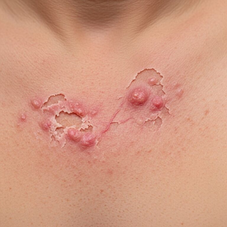
Benign Adnexal Tumours
The majority of adnexal neoplasms encountered in clinical dermatology are benign lesions that rarely progress to malignancy. These tumours typically grow slowly and pose minimal risk to patient health, though they may be removed for cosmetic reasons or diagnostic confirmation.
Benign Eccrine and Apocrine Tumours
Benign tumours arising from sweat gland tissue include numerous entities that present with varied clinical appearances and histological features. These lesions are often asymptomatic and may be discovered incidentally during skin examination or imaging studies performed for other reasons.
Benign Follicular Tumours
Tumours originating from hair follicle structures represent another significant category of benign adnexal neoplasms. These lesions demonstrate characteristic features of follicular differentiation on histological examination and typically remain localized without aggressive behavior.
Sebaceous Tumours
Benign sebaceous tumours include various lesions derived from sebaceous gland tissue. These tumours are identified through the characteristic “mulberry cell” appearance and sebaceous differentiation patterns on microscopy. Most sebaceous tumours remain benign, though rare malignant variants have been documented.
Malignant Adnexal Tumours
While most adnexal tumours are benign, malignant variants do exist and require appropriate clinical attention and management. Malignant adnexal neoplasms may originate from any of the three primary adnexal structures and generally require more aggressive treatment than their benign counterparts.
Malignant Eccrine and Apocrine Tumours
Malignant tumours of sweat gland origin include several aggressive entities that warrant careful evaluation and prompt treatment. These tumours may demonstrate rapid growth, infiltrative behavior, and potential for metastasis.
Malignant Follicular Tumours
Malignant tumours derived from hair follicle structures represent a less common but important category of cutaneous malignancies. One notable example is trichoblastic carcinoma, a rare malignant variant that may arise from trichoblastoma precursor lesions.
Trichoblastic carcinoma differs from classic basal cell carcinoma (BCC) in several important ways. While many authors consider trichoblastic carcinoma as synonymous with BCC based on current understanding of BCC as a tumour of skin adnexal structures, the rare trichoblastic carcinoma arising in trichoblastoma demonstrates a notably higher potential for distant metastasis compared to typical BCC. This variant is characterized by a “bottom heavy” configuration, restricted tumour stroma, and may infiltrate into underlying muscles, underscoring its more aggressive nature.
Diagnostic Approach and Evaluation
When a suspected adnexal tumour is identified, a systematic diagnostic approach ensures accurate classification and appropriate management. Clinical evaluation should be combined with histopathological examination for definitive diagnosis.
Clinical Assessment
Initial evaluation includes careful examination of the lesion’s size, location, appearance, and associated symptoms. However, clinical features alone are insufficient for definitive diagnosis, as many adnexal tumours present with non-specific appearances.
Biopsy and Histopathology
Tissue biopsy remains the gold standard for definitive diagnosis of adnexal tumours. Biopsy techniques may include punch biopsy, shave biopsy, or excisional biopsy, depending on the clinical context and suspected diagnosis. Specimens must be properly fixed and processed to allow for adequate histological evaluation.
Special Stains and Immunohistochemistry
Beyond routine H&E staining, special histochemical stains and immunohistochemical markers may be employed to confirm the specific type of adnexal differentiation and support diagnostic accuracy. These adjunctive techniques can clarify ambiguous cases and provide additional prognostic information.
Management and Treatment Considerations
Management of adnexal tumours depends on multiple factors, including whether the lesion is benign or malignant, its size, location, and the patient’s clinical presentation and preferences.
Benign Lesions
Most benign adnexal tumours require no treatment beyond diagnostic confirmation through biopsy. However, lesions that are cosmetically concerning, symptomatic, or located in areas subject to trauma may be removed surgically. Excision typically provides complete resolution with excellent cosmetic outcomes when performed appropriately.
Malignant Lesions
Malignant adnexal tumours generally require more aggressive treatment strategies. Surgical excision with adequate margins represents the primary treatment approach for most malignant adnexal neoplasms. Additional considerations may include:
- Wide local excision with histologically confirmed clear margins
- Mohs micrographic surgery for certain lesions requiring margin verification
- Lymph node evaluation and staging for tumours with metastatic potential
- Adjuvant therapies such as radiation or chemotherapy for high-risk lesions
Specific Tumour Categories
Hair Follicle-Derived Tumours
Tumours demonstrating follicular differentiation include both benign entities such as trichofolliculomas and pilomatricomas, as well as malignant variants including follicular carcinomas. These tumours exhibit characteristic histological features of hair follicle differentiation on microscopic examination.
Sebaceous Gland-Derived Tumours
Sebaceous tumours constitute a diverse group including sebaceous adenomas, sebaceomas, and sebaceous carcinomas. The identification of sebaceous differentiation relies on recognition of characteristic lipid-rich cells with vacuolated cytoplasm. While most sebaceous tumours are benign, sebaceous carcinoma represents an aggressive malignancy that may occur sporadically or in association with Muir-Torre syndrome.
Sweat Gland-Derived Tumours
Eccrine and apocrine sweat gland tumours represent a large category of adnexal neoplasms. Benign lesions include syringomas, eccrine poroma, and hidradenomas, while malignant variants include eccrine carcinoma and malignant apocrine neoplasms. These tumours demonstrate characteristic ductal structures and secretory differentiation on histological examination.
Clinical Significance and Prognosis
Understanding the benign versus malignant nature of adnexal tumours is critical for appropriate patient counseling and treatment planning. The vast majority of adnexal tumours are benign and carry excellent prognoses following diagnosis and appropriate management.
Malignant adnexal tumours, while uncommon, require careful attention and appropriate treatment to prevent progression and metastatic disease. Early recognition and appropriate therapy improve outcomes in these cases. Patients with malignant adnexal neoplasms may require long-term follow-up to monitor for recurrence or metastatic disease.
Frequently Asked Questions
Q: Are adnexal tumours cancer?
A: Most adnexal tumours are benign and not cancerous. However, some malignant variants do exist, which is why definitive diagnosis through biopsy and histopathological examination is essential.
Q: How are adnexal tumours diagnosed?
A: Diagnosis relies primarily on histopathological examination of tissue obtained through biopsy, rather than clinical appearance alone. Special stains and immunohistochemistry may support diagnosis in complex cases.
Q: Do benign adnexal tumours require treatment?
A: Most benign adnexal tumours require no treatment beyond diagnostic confirmation. Surgical removal may be considered for cosmetic reasons or if the lesion is symptomatic or located in an area subject to trauma.
Q: What is the treatment for malignant adnexal tumours?
A: Malignant adnexal tumours typically require surgical excision with adequate margins. Some cases may benefit from additional therapies such as radiation or chemotherapy, particularly for high-risk lesions.
Q: Can adnexal tumours recur after treatment?
A: Benign adnexal tumours rarely recur after complete surgical excision. Malignant tumours have a higher recurrence risk, and long-term follow-up is typically recommended to monitor for recurrence or metastatic disease.
Q: What is the difference between benign and malignant adnexal tumours?
A: Benign adnexal tumours grow slowly and rarely progress to malignancy, while malignant variants may demonstrate rapid growth, infiltrative behavior, and potential for metastasis. Histopathological features help distinguish between these categories.
References
- Skin adnexal neoplasms—part 1: An approach to tumours of the hair follicle, sebaceous, eccrine and apocrine units — National Center for Biotechnology Information (NCBI/PMC). 2008. https://pmc.ncbi.nlm.nih.gov/articles/PMC1860623/
- Adnexal tumour — DermNet New Zealand. Updated 2025. https://dermnetnz.org/topics/adnexal-tumour
- Benign Adnexal Masses — Merck Manuals Professional Edition. 2024. https://www.merckmanuals.com/professional/gynecology-and-obstetrics/miscellaneous-gynecologic-disorders/benign-adnexal-masses
Similar Articles


Read full bio of Sneha Tete