Airborne Contact Dermatitis: 5 Types And Top Treatments
Understanding skin inflammation from airborne particles: causes, symptoms, diagnosis, and effective management strategies.

Airborne contact dermatitis is an acute or chronic inflammatory skin condition primarily affecting exposed body areas, especially the face, due to particles suspended in the air such as fibres, dust, vapours, sprays, gases, and plant materials.
Introduction
Contact dermatitis is classified as airborne when skin inflammation results from airborne agents rather than direct touch. This form spares covered areas like the eyelids less prominently than direct contact types but characteristically involves photo-exposed sites symmetrically. It poses diagnostic challenges due to diverse sources and presentations.
Patients often report sudden onset after environmental changes, occupational shifts, or seasonal exposures. Early recognition prevents chronicity, as persistent exposure can lead to lichenification or secondary infections.
Classification
Airborne contact dermatitis encompasses several subtypes based on mechanism:
- Airborne irritant contact dermatitis: From non-immunologic direct tissue damage by substances like dust or gases.
- Airborne allergic contact dermatitis: Type IV delayed hypersensitivity to haptens penetrating the skin.
- Airborne phototoxic reactions: Non-immunologic responses triggered by light exposure on chemicals like psoralens.
- Airborne photoallergic reactions: Immunologic responses combining allergen and UV light, e.g., from certain fragrances.
- Airborne contact urticaria: Immediate type I hypersensitivity causing wheals, from proteins like pollen.
These classifications guide testing and management, with allergic forms requiring patch tests and photoallergic needing photopatch series.
Demographics
Affected individuals span all ages but commonly include outdoor workers in agriculture, construction, painting, and horticulture. Atopics with prior eczema history show heightened susceptibility due to barrier defects. Women may encounter non-occupational sources via cosmetics or plants. In endemic areas like India, Parthenium hysterophorus weed affects thousands annually, predominantly farmers.
Occupational clusters appear in healthcare (disinfectants), manufacturing (resins), and floristry (pollens). Global reports highlight regional variations, e.g., sesquiterpene lactones in Europe from Compositae plants.
Causes
Sources divide into occupational and non-occupational. Diagnosis as airborne relies on:
- Volatile or airborne agent presence.
- Symmetrical rash on exposed sites.
- History of exposure without direct contact.
- Positive epicutaneous tests.
Airborne allergens (allergic contact dermatitis):
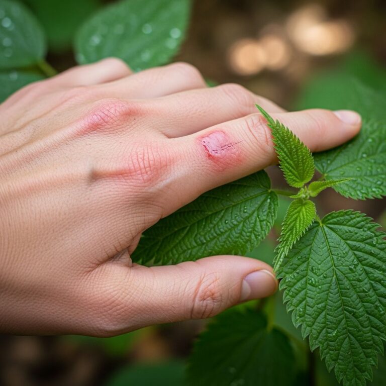
- Plants: Parthenium hysterophorus, ragweed, chrysanthemum (sesquiterpene lactones).
- Chemicals: Epoxy resins, acrylates, formaldehyde resins in textiles.
- Metals: Cobalt, chromate dust.
- Preservatives: Isothiazolinones in paints, Thiomersal.
- Fragrances: Balsam of Peru volatiles.
Airborne irritants (irritant contact dermatitis):
- Fibreglass, rock wool.
- Pesticides, solvents.
- Ammonium persulfate (hairdressing).
- Phosphorus sesquisulfide (match industry).
Photoallergic/photoaggravated:
- Phenothiazines, musk ambrette.
- 6-Methylcoumarin, ketoprofen.
Contact urticaria agents:
- Pollen grains, insect debris.
- Latex proteins, food powders.
Immunologically, allergic forms involve hapten-protein conjugation, Langerhans cell migration to lymph nodes, T-cell priming, and effector recall on re-exposure, yielding inflammation via cytokines.
Clinical Features
Symptoms include intense itching, burning, or stinging, worse outdoors or at work. Rash appears symmetrically on face (forehead, eyelids, cheeks sparing nasolabial folds), neck, V-neck chest, dorsal forearms/hands, sparing flexures and palms.
Morphology: Diffuse scaly erythematous macules or plaques; acute weeping vesicles; chronic lichenified thickening. Eyelids show marked edema. Secondary pustules arise from bacterial superinfection. Rare variants: urticarial, acneiform, lichenoid, erythema multiforme-like, purpuric, or pigmentary.
| Site | Typical Features |
|---|---|
| Face | Symmetric erythema, scaling; eyelid edema |
| Neck/Chest | Well-demarcated V-area |
| Forearms | Dorsal sparing volar |
| Rare | Scalp, anogenital (settling particles) |
Photodistribution predominates in photoallergic cases. Parthenium dermatitis spreads to covered sites via self-transfer.
Diagnosis
Challenging due to mimicry; hinges on history (exposure timeline, improvement indoors), exam (exposed-site pattern), and tests. Key differentials: seborrheic dermatitis (nasolabial involvement), airborne photocontact, atopic dermatitis (flexural), lupus (malar rash).
- Patch testing: Baseline series plus occupational/environmental suspects; positive if airborne-relevant concentration reproduces pattern.
- Photopatch testing: For suspected photoallergens.
- Prick testing: Urticaria suspects.
- Histology: Spongiosis, lymphocytic infiltrate; nonspecific.
Open testing or use tests simulate airborne exposure. Phototesting rules out actinic dermatitis.
Differential Diagnoses
| Condition | Key Distinguishers |
|---|---|
| Atopic dermatitis | Flexural, personal/family atopy history, no patch positives |
| Seborrhoeic dermatitis | Greasy scales in nasolabial folds, scalp |
| Systemic contact dermatitis | Generalized, ingested allergen |
| Photodermatitis | Strict photo-distribution, minimal eyelids |
| Cosmetic dermatitis | Asymmetric, direct product application |
Treatment
Tailored to cause; cornerstone is avoidance via relocation, ventilation, PPE (masks, goggles). Occupational counseling essential.
Mild-moderate:
- Cool wet compresses 15-30 min several times daily.
- Emollients frequently.
- Topical corticosteroids (potent for face: methylprednisolone aceponate).
Severe/extensive:
- Oral prednisolone 0.5-1 mg/kg tapered over 3 weeks.
- Phototherapy (narrowband UVB, PUVA) for chronic photoallergic.
- Immunosuppressants: Azathioprine (1.5-2.5 mg/kg), methotrexate (15 mg/week) for recalcitrant cases like Parthenium.
Antibiotics for pustules. Barrier creams prevent irritant penetration.
Outcome
Prognosis excels with exposure cessation; full recovery typical. Chronic cases risk persistent actinic dermatitis or erythroderma. Quality-of-life impact significant from disfigurement, itch. Patient education on allergen avoidance critical; some require career shifts.
Frequently Asked Questions (FAQs)
What causes airborne contact dermatitis?
Airborne particles like plant pollen, chemical dusts, fibreglass, or vapours from paints and pesticides trigger it on exposed skin.
How is airborne contact dermatitis diagnosed?
Through clinical history, rash pattern on exposed areas, and confirmatory patch or photopatch testing.
Can airborne contact dermatitis affect covered skin?
Rarely, via particle settling under clothing or self-transfer, especially with oily allergens.
What is the best treatment for airborne contact dermatitis?
Avoidance of the trigger, combined with topical steroids and emollients; severe cases need systemic therapy.
Is airborne contact dermatitis contagious?
No, it is not infectious; it results from individual sensitivity to environmental agents.
References
- Airborne contact dermatitis – Dr. Breslavets | CMSD — Canadian Medical Skin Dermatology. 2023. https://cmsderm.ca/airborne-contact-dermatitis/
- Airborne contact dermatitis – DermNet — DermNet NZ. 2024-01-15. https://dermnetnz.org/topics/airborne-contact-dermatitis
- AIRBORNE CONTACT DERMATITIS – CURRENT PERSPECTIVES — PMC / NCBI. 2012-01-27. https://pmc.ncbi.nlm.nih.gov/articles/PMC3276900/
- Contact dermatitis – Diagnosis and treatment – Mayo Clinic — Mayo Clinic. 2025-06-10. https://www.mayoclinic.org/diseases-conditions/contact-dermatitis/diagnosis-treatment/drc-20352748
Similar Articles


Read full bio of Sneha Tete