Amelanotic Melanoma: Why It Doesn’t Look Like Other Melanomas
Learn why amelanotic melanoma is harder to detect and what warning signs to watch for.

Amelanotic Melanoma: It Doesn’t Look Like Other Melanomas
Melanoma is one of the most dangerous forms of skin cancer, and early detection is crucial for improving survival rates. However, not all melanomas look the same. Amelanotic melanoma is a rare subtype that presents unique diagnostic challenges because it lacks the dark pigmentation typically associated with melanoma. Understanding this disease and recognizing its atypical features can mean the difference between early treatment and advanced cancer.
What Is Amelanotic Melanoma?
Amelanotic melanoma (AM) is a subtype of melanoma characterized by little to no melanin pigmentation. The term “amelanotic” refers to the absence of pigment, which is what makes this form of melanoma distinctly different from typical melanomas. Amelanotic melanomas account for less than 2% of all melanoma diagnoses, making them relatively uncommon. However, despite their rarity, these lesions are associated with higher mortality rates compared to pigmented melanomas.
The primary challenge with amelanotic melanoma is that patients and clinicians may not immediately recognize it as a melanoma. Without the characteristic dark color, these lesions can be mistaken for benign skin conditions, basal cell carcinoma, squamous cell carcinoma, or other non-cancerous growths. This misdiagnosis can lead to dangerous delays in treatment and more advanced disease at the time of diagnosis.
Why Amelanotic Melanoma Is Hard to Diagnose
The diagnostic challenge posed by amelanotic melanoma stems from its atypical appearance. Amelanotic melanomas do not typically display the classic ABCDE warning signs that dermatologists teach patients to watch for in pigmented melanomas. These traditional signs include asymmetry, border irregularity, color variation, diameter greater than 6 millimeters, and evolution or change over time.
Without obvious pigmentation, patients and healthcare providers may fail to recognize these lesions as suspicious. Studies have shown that amelanotic melanomas are often more advanced at the time of diagnosis compared to pigmented melanomas, meaning they have progressed to a later stage. This delay in diagnosis directly impacts treatment options and patient outcomes.
Clinical Appearance of Amelanotic Melanoma
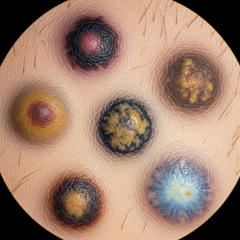
One of the defining characteristics of amelanotic melanoma is its appearance. Rather than the dark brown or black color associated with typical melanomas, amelanotic lesions may present in several ways:
- Skin-colored: The lesion may appear similar in color to surrounding normal skin
- Pink or red: Many amelanotic melanomas present as pink, red, or erythematous lesions
- Flesh-colored papules: Some appear as raised, flesh-colored bumps or nodules
- With faint pigmentation: Some lesions may have light tan, brown, or gray pigmentation at the periphery
Early amelanotic melanomas typically present as asymmetrical macular lesions that are uniformly pink or red. The borders may be either well-defined or ill-defined, adding to the diagnostic confusion. A significant proportion of amelanotic melanomas also display a scaly appearance, which may be related to increased keratinocyte turnover.
Expanding Warning Signs: The 3 Rs
Because amelanotic melanomas do not always follow the classic ABCDE criteria, dermatologists have expanded the warning signs to include additional features. The 3 Rs supplement the traditional ABCD criteria:
- Red: The lesion appears red or pink rather than brown or black
- Raised: The lesion is elevated or has a nodular appearance
- Recent change: The lesion has changed in size, shape, or appearance over weeks or months
Additionally, some experts use the EFG criteria for amelanotic melanoma detection:
- Elevated: The lesion stands out above the surrounding skin
- Firm: The lesion feels firm to the touch
- Growing: The lesion has been enlarging for more than one month
Patients and clinicians should be alert to any new or changing skin lesion, particularly if it meets any of these criteria.
Dermoscopy and Vessel Morphology
Dermoscopy, the examination of skin lesions using a specialized magnifying instrument called a dermatoscope, is a vital diagnostic tool for detecting amelanotic melanoma. When melanin pigmentation is absent or minimal, the focus shifts to evaluating other features of the lesion.
Vascular morphology—the pattern and structure of blood vessels within the lesion—is the most important dermoscopic feature for identifying suspicious amelanotic melanomas. Abnormal vessel patterns raise suspicion for melanoma and include:
- Irregular dot vessels
- Linear irregular vessels
- Arborising (branching) vessels
- Polymorphic (varied) vessels
The 5 + 2 list is recommended as a guide for vessel analysis in lesions lacking pigmentation. Different vessel morphologies may indicate disease progression. Dotted vessels that are homogenous in shape are associated with early, flat amelanotic melanomas. As the tumor advances, linear vessels appear in increasing numbers. More advanced disease shows longer, coarser vessels that vary in shape.
Interestingly, “comma-like” vessel morphology has been found to be a significant negative indicator for amelanotic melanoma, meaning its presence suggests the lesion is less likely to be melanoma. The distribution of vasculature is also important—atypical vessel distribution along the peripheral edges of the skin lesion supports a diagnosis of amelanotic melanoma.
Risk Factors for Amelanotic Melanoma
Several factors increase the risk of developing amelanotic melanoma, similar to the risk factors for other types of melanoma:
- Personal or family history of melanoma
- History of five or more blistering sunburns before age 20
- Presence of more than 50 benign moles
- Five or more atypical moles with large size, color variations, or indistinct borders
- Fair skin that does not tan easily and frequently burns
- Skin with many freckles or tendency to freckle after sun exposure
- Blue or green eyes
- Naturally red or blonde hair
Individuals with these risk factors should be especially vigilant about monitoring their skin and seeking regular dermatological evaluation.
Diagnostic Procedures
Diagnosis of amelanotic melanoma requires a biopsy, the gold standard for confirming skin cancer. The diagnostic process typically involves:
- Clinical examination: A dermatologist examines the lesion clinically and with dermoscopy
- Local anesthesia: The area is numbed with local anesthetic
- Tissue removal: The suspicious lesion is carefully removed, ideally with a 2-3 millimeter clinical margin
- Pathological examination: A dermatopathologist examines the tissue microscopically
- Pathology report: The report documents findings including depth of invasion, Clark level, and Breslow thickness
The pathology report includes critical prognostic information. Breslow thickness—the depth of tumor invasion measured in millimeters—is the most important prognostic factor. Clark level indicates the anatomical plane of invasion and is particularly useful for predicting outcomes in thin tumors.
Prognosis and Outcomes
The prognosis of amelanotic melanoma is similar to that of pigmented melanomas when matched for stage and depth. However, because amelanotic melanomas are often diagnosed at a more advanced stage due to delayed recognition, overall outcomes may be worse. Studies show that amelanotic melanoma has a greater hazard of death compared to pigmented melanoma, with a hazard ratio of 2.0.
Key prognostic factors include:
- Breslow thickness: The most important factor; thinner tumors have better prognosis
- Location of the lesion: Anatomical location affects outcomes
- Patient age: Age influences prognosis
- Patient sex: Sex may influence disease progression
Distinguishing Amelanotic Melanoma From Other Conditions
Amelanotic melanoma can mimic numerous other skin conditions, which contributes to diagnostic delays. Common conditions that amelanotic melanoma may resemble include:
- Basal cell carcinoma
- Squamous cell carcinoma
- Benign moles or nevi
- Skin cysts
- Scars
- Erythematous plaques
- Nodular lesions
This is why complete skin examination is essential. Examining the entire skin surface for sun damage (such as actinic keratoses) and other pigmented lesions provides clinical context that may raise suspicion for a non-pigmented lesion being amelanotic melanoma.
Clinical Characteristics and Demographics
Studies examining amelanotic melanoma characteristics have revealed important epidemiological patterns. In one retrospective analysis, 68.75% of amelanotic melanoma patients were male, with a mean age at diagnosis of 78 years. Amelanotic melanomas often present as the nodular subtype, which is known for aggressive vertical growth—a characteristic that contributes to more advanced disease at diagnosis.
Histopathologically, approximately 8% of melanomas are amelanotic. When melanomas are identified as amelanotic, they generally present at a higher tumor stage compared to pigmented melanomas, highlighting the importance of heightened clinical awareness.
Why Patient History Matters
A patient’s history of observing change in a lesion is an important diagnostic factor, particularly for amelanotic lesions. Unlike pigmented melanomas, where obvious color change may prompt earlier concern, amelanotic melanomas may change in ways that are less visually obvious. Patients should be educated to report any changes in existing skin lesions, including:
- Changes in size (growth)
- Changes in shape (becoming irregular)
- Changes in texture (becoming scaly or rough)
- Appearance of new bumps or raised areas
- Any bleeding or oozing
- Any itching or tenderness
Importance of Vigilance in Clinical Practice
Healthcare practitioners in both primary and secondary care settings must be aware of amelanotic melanoma and vigilant in identifying it. The significant misdiagnosis rate associated with amelanotic melanoma—combined with its potential for aggressive progression—makes heightened clinical awareness essential. Key recommendations for clinicians include:
- Maintain high clinical suspicion for non-pigmented lesions in at-risk patients
- Use dermoscopy to evaluate vascular morphology in lesions lacking pigmentation
- Refer suspicious lesions for excision biopsy with appropriate margins
- Educate patients about the expanded warning signs (including the 3 Rs)
- Consider amelanotic melanoma in the differential diagnosis of any changing skin lesion
Frequently Asked Questions
Q: What is the difference between amelanotic melanoma and regular melanoma?
A: Amelanotic melanoma lacks melanin pigmentation, appearing skin-colored, pink, or red instead of the dark brown or black typical of regular melanomas. This makes it harder to recognize and is often diagnosed at a more advanced stage.
Q: How common is amelanotic melanoma?
A: Amelanotic melanoma accounts for less than 2% of all melanoma diagnoses, making it a relatively rare subtype despite its aggressive behavior.
Q: Why is amelanotic melanoma so dangerous?
A: Amelanotic melanoma is dangerous because it often goes unrecognized, leading to delayed diagnosis and more advanced disease at the time of treatment. Studies show it has a higher mortality rate compared to pigmented melanomas.
Q: What should I do if I notice a changing skin lesion?
A: Any new or changing skin lesion, particularly one that is red, pink, raised, or growing, should be evaluated by a dermatologist. This is especially important if the lesion meets any of the ABCDEs or 3 Rs criteria.
Q: Can dermoscopy definitively diagnose amelanotic melanoma?
A: While dermoscopy is helpful for identifying suspicious features—particularly abnormal vessel patterns—only a biopsy can definitively diagnose amelanotic melanoma. Any suspicious lesion should be biopsied.
Q: What does Breslow thickness mean?
A: Breslow thickness is the depth of melanoma invasion measured in millimeters. It is the most important factor in determining prognosis, with thinner tumors having better outcomes.
References
- Identifying the clinical and histopathological characteristics of amelanotic melanoma — National Center for Biotechnology Information (NIH/NLM). 2024. https://pmc.ncbi.nlm.nih.gov/articles/PMC11049562/
- Amelanotic melanoma: Appearances, Symptoms and Treatment — DermNet New Zealand. 2024. https://dermnetnz.org/topics/amelanotic-melanoma
- Amelanotic Melanoma Overview — Moffitt Cancer Center. 2024. https://www.moffitt.org/cancers/melanoma/diagnosis/types/amelanotic-melanoma/
- Amelanotic melanoma: a unique case study and review of the literature — Western Pathology Inc. 2018. https://westernpathologyinc.com/site/wp-content/uploads/2020/06/2018-Amelanotic-melanoma-a-unique-case-study-and-review-of-the-literature-Bruce-Ragsdale-MD.pdf
- Amelanotic Melanoma: Symptoms, Stages & Growth Rate — Cancer Center. 2024. https://www.cancercenter.com/cancer-types/melanoma/types/amelanotic-melanoma
- How to recognise amelanotic melanoma — Pulse Today. 2024. https://www.pulsetoday.co.uk/clinical-feature/clinical-areas/dermatology-and-wound-care/how-to-recognise-amelanotic-melanoma/
Similar Articles


Read full bio of medha deb