Angiofibroma: Types, Appearances and Causes
Comprehensive guide to angiofibromas: benign vascular skin lesions and their clinical management.

Introduction
An angiofibroma (AGF) is a benign vascular neoplasm composed of dermal fibrous tissue and blood vessels. These lesions represent a diverse group of cutaneous growths that vary in their clinical presentation, etiology, and association with systemic genetic disorders. Angiofibromas are characterized by their benign nature, meaning they do not undergo malignant transformation or metastasize to other body sites. The term “angiofibroma” encompasses several distinct clinical entities that can be classified based on their association with genetic syndromes or their anatomical location.
Classification and Demographics
Angiofibromas are classified into two primary categories: those associated with genetic disorders and those that are acquired without genetic predisposition. The acquired form of angiofibroma is significantly more common than syndromic presentations. These benign growths affect individuals across various age groups, though specific types show predilection for particular demographics. For instance, juvenile nasopharyngeal angiofibromas (JNA) almost exclusively affect teenage boys, typically between ages 7 and 19, with peak diagnosis occurring between ages 10 and 19. Geographic variations in prevalence have been documented, with higher incidence rates reported in Egypt, India, Southeast Asia, Kenya, and Mexico compared to other regions.
Genetic Associations
Several hereditary syndromes are associated with angiofibroma development:
Tuberous Sclerosis Complex
Tuberous sclerosis is a neurocutaneous autosomal dominant syndrome in which angiofibromas appear during childhood, predominantly in the nasolabial folds and on the central face. These facial lesions were historically termed “adenoma sebaceum,” “juvenile angiofibroma,” or “Pringle tumour,” though current nomenclature favors the term angiofibroma. Patients with tuberous sclerosis frequently develop additional cutaneous manifestations, including periungual angiofibromas (Koenen tumours) and oral fibromas over time. The condition results from mutations in the TSC1 or TSC2 genes, which encode proteins that regulate cell growth and division, affecting the skin, brain, kidneys, heart, and lungs.
Birt-Hogg-Dubé Syndrome
This rare genetic disorder causes skin and kidney tumors, lung cysts, and pneumothorax. The condition results from mutations in the FLCN gene and presents with angiofibromas as one of several characteristic skin manifestations, along with fibrofolliculomas and trichodiscomas.
Multiple Endocrine Neoplasia Type 1
This hereditary syndrome leads to tumors in multiple endocrine organs, including the parathyroid glands, pituitary gland, and pancreas. Mutations in the MEN1 gene result in loss of tumor suppressor function, and angiofibromas represent one of the cutaneous signs alongside collagenomas and lipomas.
Acquired Angiofibromas
Acquired angiofibromas develop spontaneously or through unknown mechanisms without association with genetic syndromes. These lesions can appear on various body sites including the nose, face, penis, and mouth. They are composed of collagen, fibroblasts, and blood vessels, representing benign growths that occur independently of systemic disease.
Clinical Features and Appearances
Cutaneous Presentations
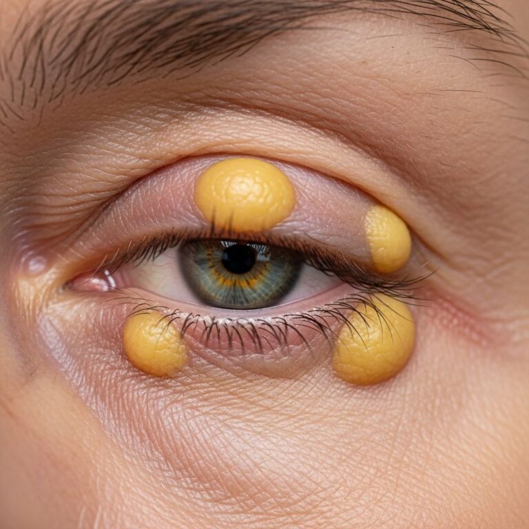
Angiofibromas associated with tuberous sclerosis typically appear as small, red bumps on the face, especially on the nose and cheeks. These lesions characteristically manifest in childhood and may enlarge slowly throughout adolescence and early adulthood, causing significant textural changes to the skin. A fibrous papule, considered an acquired form of angiofibroma, is characteristically found in adults as a solitary lesion usually on the nose, and is often clinically mistaken for basal cell carcinoma or melanocytic naevus.
Nasopharyngeal Presentations
Juvenile nasopharyngeal angiofibromas present with distinct clinical features related to their anatomical location in the nasal cavity and nasopharynx. These lesions typically appear as red, raised bumps that may bleed upon contact. The tumors generally grow through adolescence and into the early 20s, with some spontaneous regression occurring after late teens. Approximately one in five cases develop after age 20, and cases have been documented in men up to age 50, though almost all patients are male.
Symptoms and Complications
Primary Symptoms
The most common symptoms of angiofibroma vary depending on anatomical location and tumor size:
- Nasal obstruction or difficulty breathing through the nose
- Frequent or repeated nosebleeds, often from a single nostril
- Nasal discharge, typically blood-tinged
- Persistent runny nose unresponsive to standard treatment
- Headaches and facial pain, particularly in sinus regions
- Facial swelling, including cheek enlargement
- Stuffy or congested nose
Advanced Symptoms from Nerve and Tissue Compression
As angiofibromas enlarge, they may compress important anatomical structures, leading to additional complications:
- Hearing loss resulting from tumor extension into the eustachian tube
- Vision changes, double vision, or blindness from optic nerve damage
- Loss of sense of smell (anosmia)
- Facial abnormalities including drooping eyelids and bulging eyes
- Numbness or altered sensation in the face
Serious Complications
Untreated angiofibromas may continue growing, with symptoms progressively worsening. Potential serious complications include life-threatening nosebleeds that require emergency intervention, bone damage from tumor invasion into surrounding structures, and permanent loss of sensory or motor function from chronic nerve compression. The prognosis remains benign in terms of malignant transformation, as angiofibromas do not transform into cancerous lesions. However, they may persist or recur following treatment, and complications can arise from both the tumor itself and associated genetic disorders that may involve other organ systems.
Diagnosis and Clinical Evaluation
Angiofibromas are diagnosed through clinical appearance and confirmed by histopathology. The diagnostic approach typically involves:
- Clinical examination assessing the characteristic appearance and location of lesions
- Skin biopsy to examine tissue under microscopy and rule out differential diagnoses
- Imaging studies (CT or MRI) for nasopharyngeal lesions to assess tumor size, extent, and relationship to surrounding structures
- Endoscopic evaluation for intranasal tumors to visualize the lesion directly
- Genetic testing when syndromic angiofibroma is suspected to identify underlying genetic disorders
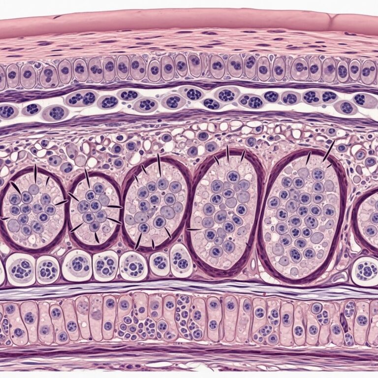
Histopathological examination reveals a benign lesion composed of fibrovascular tissue with characteristic features distinguishing it from other cutaneous and mucosal lesions.
Differential Diagnoses
Several conditions may resemble angiofibromas clinically and must be excluded during the diagnostic process. Fibrous papules on the nose can be mistaken for basal cell carcinoma or melanocytic naevi, necessitating biopsy confirmation. Nasopharyngeal angiofibromas must be differentiated from other nasal masses including nasal polyps, hemangiomas, and other vascular lesions. The presence of syndromic features and family history helps distinguish genetic syndrome-associated angiofibromas from acquired forms.
Treatment Options
Treatment approaches for angiofibromas depend on the specific type, location, size, and functional impact of the lesion:
Observation
Many acquired cutaneous angiofibromas remain stable and may not require treatment unless they cause cosmetic or functional concerns. Observation with regular monitoring allows assessment of growth patterns and development of symptoms.
Surgical Intervention
Surgical excision represents the primary treatment for symptomatic nasopharyngeal angiofibromas and cutaneous lesions causing significant cosmetic or functional impairment. Complete surgical removal is the goal to minimize recurrence risk.
Cosmetic and Dermatological Treatments
For cutaneous angiofibromas, particularly those associated with tuberous sclerosis, various dermatological interventions may improve appearance and address functional concerns, including laser therapy, cryotherapy, and topical pharmacological agents.
Prognosis and Outcomes
Angiofibromas have a benign clinical course and do not transform into malignant tumors. However, they may persist or recur after treatment, particularly if complete surgical removal is not achieved. The natural history of juvenile nasopharyngeal angiofibromas suggests growth through adolescence and early adulthood, with potential spontaneous regression after the late teens. The long-term prognosis also depends on the presence and severity of systemic involvement in associated genetic disorders.
Complications of Treatment
While angiofibromas themselves are benign and non-contagious, treatment-related complications may include scarring, post-procedural pain, infection, and recurrence. Patients should be counseled about these potential outcomes and the importance of close follow-up after treatment.
Frequently Asked Questions
Q: Are angiofibromas cancerous?
A: No, angiofibromas are benign (non-cancerous) growths that do not transform into malignant tumors or metastasize to other sites. However, they can cause significant morbidity through compression of nearby structures and should be monitored clinically.
Q: Who is most likely to develop angiofibromas?
A: Juvenile nasopharyngeal angiofibromas almost exclusively affect teenage boys, typically between ages 7 and 19. Cutaneous angiofibromas associated with tuberous sclerosis appear in childhood, while acquired angiofibromas can develop in adults of either sex.
Q: Can angiofibromas be contagious?
A: No, angiofibromas are not contagious and are not caused by infection or transmitted through contact. They are growths of blood vessels and fibrous tissue without infectious etiology.
Q: What should I do if I suspect I have an angiofibroma?
A: If you notice persistent nasal symptoms, facial lesions, or other concerning features, consult a healthcare provider for clinical evaluation. Diagnosis involves physical examination and may include biopsy or imaging studies to confirm the diagnosis and assess extent of disease.
Q: Are there genetic tests for syndromic angiofibromas?
A: Yes, genetic testing is available for tuberous sclerosis complex, Birt-Hogg-Dubé syndrome, and multiple endocrine neoplasia type 1. Genetic counseling is recommended for individuals with suspected syndromic angiofibromas to assess inheritance patterns and identify affected family members.
Q: Will my angiofibroma grow throughout my life?
A: Juvenile nasopharyngeal angiofibromas typically grow through adolescence and early adulthood, with potential spontaneous regression after the late teens. Cutaneous angiofibromas may enlarge slowly, though acquired forms often remain stable. Individual growth patterns vary, necessitating clinical monitoring.
References
- DermNet NZ – Angiofibroma — DermNet New Zealand, Dermatological Society of New Zealand. 2024. https://dermnetnz.org/topics/angiofibroma
- Juvenile Nasopharyngeal Angiofibroma — Nemours KidsHealth, Nemours Children’s Health. 2024. https://kidshealth.org/en/parents/juvenile-asopharyngeal-angiofibroma.html
- Juvenile Angiofibromas — Mount Sinai Medical Center. 2024. https://www.mountsinai.org/care/pediatrics/services/pediatric-cerebrovascular-disorders/juvenile-angiofibromas
- Angiofibromas Treatment — Porter Ranch Dermatologist. 2024. https://porterranchdermatologist.com/medical-dermatology/angiofibromas/
- Definition of Angiofibroma — National Cancer Institute Dictionary of Cancer Terms. U.S. National Institutes of Health. 2024. https://www.cancer.gov/publications/dictionaries/cancer-terms/def/angiofibroma
- Angiofibromas — Children’s Hospital of Philadelphia. 2024. https://www.chop.edu/conditions-diseases/angiofibromas
Similar Articles


Read full bio of Sneha Tete