Angiolymphoid Hyperplasia with Eosinophilia
Understanding ALHE: A rare benign skin condition with vascular proliferation, eosinophilic infiltrate, and challenging treatment options.

Author: Reviewed by Dr. Amanda Oakley, Dermatologist, Hamilton, New Zealand. DermNet NZ Editor: Dr Martin Steinhoff. Last updated October 2024 at 11:53 PM UTC.
What is angiolymphoid hyperplasia with eosinophilia?
Angiolymphoid hyperplasia with eosinophilia (ALHE), also known as epithelioid hemangioma, is a rare, benign vasoproliferative disorder characterized by a proliferation of blood vessels lined by plump epithelioid endothelial cells accompanied by a dense inflammatory infiltrate rich in lymphocytes and eosinophils. This condition manifests clinically as solitary or multiple reddish-brown papules, plaques, or nodules, predominantly affecting the head and neck region, particularly around the ears and scalp. Although non-malignant, ALHE often recurs after treatment, posing diagnostic and therapeutic challenges.
The term ALHE reflects its histopathological hallmarks: angio (vascular proliferation), lymphoid (lymphocytic infiltrate), and eosinophilia (eosinophil-rich inflammation). It primarily involves the dermis and subcutis but can extend extracutaneously to sites like the lacrimal gland, bone, or oral mucosa. ALHE is uncommon, with females more frequently affected after age 30, and lesions may be pruritic, painful, or asymptomatic.
Who gets angiolymphoid hyperplasia with eosinophilia?
ALHE typically occurs in adults aged 20–50 years, with a female predominance (approximately 2:1 ratio), though cases in children and elderly have been reported. It is rare globally, with no strong racial predilection, but has been noted in Asian populations in association with Kimura disease, though these are distinct entities. Approximately 53% of cases present as solitary lesions, while the rest are multiple or clustered.
- Prevalence: Rare; exact incidence unknown.
- Risk factors: Possible trauma (10% of cases), insect bites, or arteriovenous fistulas.
- Associations: Peripheral blood eosinophilia in 10–20% of patients; rare systemic involvement.
What causes angiolymphoid hyperplasia with eosinophilia?
The precise etiology of ALHE remains unknown, with debate centering on whether it represents a reactive inflammatory process or a true vascular neoplasm. Antigenic stimulation from insect bites or trauma is postulated in some cases (up to 10%), potentially triggering aberrant vascular proliferation. Recent immunohistochemical studies support a neoplastic origin, showing cytoplasmic WT1 staining in 95% of cases (similar to infantile hemangioma) and FOSB nuclear positivity, distinguishing it from mimics.
Other proposed factors include:
- Hyperplasia of atypical endothelial cells forming epithelioid morphology.
- Inflammatory cascade involving eosinophils and lymphocytes.
- Possible arteriovenous shunting in trauma-related cases.
No definitive genetic mutations have been identified, but the condition’s resistance to conservative therapies suggests an intrinsic proliferative drive.
What are the clinical features of angiolymphoid hyperplasia with eosinophilia?
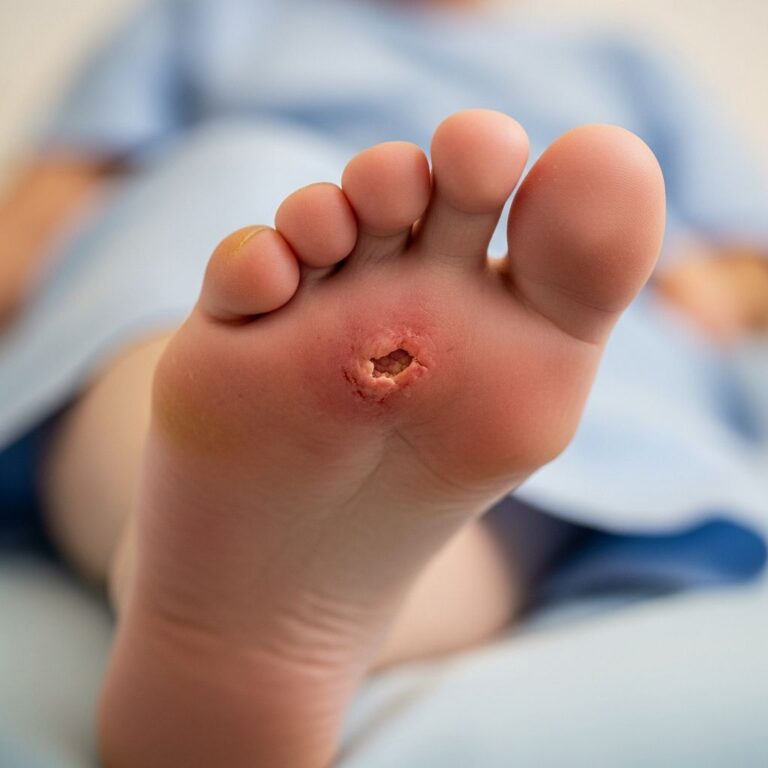
Lesions typically present as 1–2 cm, firm, reddish-brown to violaceous papules, nodules, or plaques on the head and neck (88% of cases), especially periauricular areas, scalp, and face. They may be solitary (53%) or clustered, with smooth, shiny surfaces; some exhibit ulceration or crusting from trauma. Symptoms include pruritus (common), pain, tenderness, or bleeding; lesions grow slowly over months.
| Feature | Description |
|---|---|
| Appearance | Smooth, dome-shaped papulonodules; pink/red-violet; vascular puncta visible |
| Size | 2 mm to 2 cm diameter |
| Symptoms | Itchy, tender; occasional pulsation |
| Distribution | Head/neck (88%); rare elsewhere (trunk, extremities, mucosa) |
Extracutaneous sites include lacrimal gland (proptosis), bone, colon, and vessels. Peripheral eosinophilia occurs in 10–20%; unlike Kimura disease, no lymphadenopathy.
How is angiolymphoid hyperplasia with eosinophilia diagnosed?
Diagnosis relies on clinicopathological correlation via skin biopsy, as clinical features overlap with other vascular or inflammatory lesions. Blood tests may reveal eosinophilia (10–20%), but are non-specific. Imaging (ultrasound/Doppler) can identify arteriovenous fistulas in trauma-related cases.
Pathology
Histology shows dermal/subcutaneous proliferation of irregular, thin-walled vessels lined by large epithelioid endothelial cells with abundant eosinophilic cytoplasm and vesicular nuclei. A prominent perivascular infiltrate of lymphocytes, plasma cells, and eosinophils is present; microabscesses may form. Stroma is fibrous/myxoid; epidermis often acanthotic. Immunohistochemistry: CD31+, ERG+, FOSB+, WT1+ (cytoplasmic).
- Key features: Epithelioid endothelium, eosinophilic infiltrate, no atypia.
- Differential stains: Positive for vascular markers; excludes malignancy.
Differential diagnosis for angiolymphoid hyperplasia with eosinophilia
ALHE mimics include:
| Condition | Key Differences from ALHE |
|---|---|
| Kimura disease | Deeper subcutaneous masses, lymphadenopathy, systemic eosinophilia/IgE elevation, no epithelioid vessels; Asian males |
| Epithelioid hemangioendothelioma | Malignant; atypia, mitoses, deeper invasion |
| Pyogenic granuloma | Ulcerated, lobular capillary; rapid growth, less eosinophils |
| Angiosarcoma | Atypia, dissection of collagen; elderly |
| Insect bite reaction | Superficial, resolves spontaneously; fewer vessels |
Angiolymphoid hyperplasia with eosinophilia treatment
No uniformly curative treatment exists; observation is reasonable for small lesions (3–6 months for possible regression). Surgical excision is first-line but recurs in 40–60%. Mohs micrographic surgery (MMS) offers higher cure rates by clearing deep vessels.
- Surgical: Excision, curettage, MMS (preferred for face).
- Medical: Intralesional steroids (e.g., triamcinolone 2.5–10 mg/mL; temporary response).
- Other: Pulsed dye laser, CO2 laser, cryotherapy, electrodessication, IFN-α, imiquimod; variable success.
- Emerging: Address underlying fistula if present.
Bleeding is common during surgery due to vascularity. Recurrence is frequent regardless of modality.
Frequently asked questions about angiolymphoid hyperplasia with eosinophilia
What is the prognosis for ALHE?
Benign with no malignant potential; chronic, recurrent, but self-limited in some cases.
Is ALHE contagious or hereditary?
No; not infectious or genetic.
How can I prevent ALHE recurrence?
Complete excision/MMS; avoid trauma; no proven prevention.
Does ALHE cause eosinophilia?
In 10–20%; usually mild and peripheral.
Is Kimura disease the same as ALHE?
No; deeper lesions, systemic features.
References
- Angiolymphoid Hyperplasia with Eosinophilia: Many Syllables … — Journal of Clinical and Aesthetic Dermatology (JCAD). 2019-01-01. https://jcadonline.com/angiolymphoid-hyperplasia-with-eosinophilia/
- Angiolymphoid Hyperplasia with Eosinophilia — Perri Dermatology. 2023-01-01. https://perridermatology.com/dr-perris-blog/cutaneous-vascular-anomalies-angiolymphoid-hyperplasia-with-eosinophilia/
- Epithelioid hemangioma (angiolymphoid hyperplasia with eosinophilia — YouTube (Pathology Dermatology Video). 2022-01-01. https://www.youtube.com/watch?v=zMFc0xxqTRs
- Angiolymphoid hyperplasia with eosinophilia — DermNet NZ (Official Dermatology Resource). 2024-10-01. https://dermnetnz.org/topics/angiolymphoid-hyperplasia-with-eosinophilia
- Angiolymphoid Hyperplasia With Eosinophilia — StatPearls, NCBI Bookshelf (Peer-reviewed). 2023-07-17. https://www.ncbi.nlm.nih.gov/books/NBK538181/
- Role of Eosinophils in Angiolymphoid Hyperplasia With Eosinophilia — JAMA Dermatology (Peer-reviewed). 2022-01-01. https://jamanetwork.com/journals/jamadermatology/fullarticle/2782887
Similar Articles


Read full bio of medha deb