Aponeurosis: Structure, Function, and Importance
Understanding aponeurosis: The connective tissue sheets that enable movement and support.

What Is Aponeurosis?
An aponeurosis is a thin, flat sheet of connective tissue that plays a crucial role in the structure and function of your musculoskeletal system. The term “aponeurosis” comes from Greek origins and describes a specialized fibrous tissue that serves as an anchor between your muscles and bones. Unlike tendons, which are thick, cord-like structures, aponeuroses are broad, sheet-like formations that cover muscles and provide insertion points for muscle fibers. These connective tissue structures are found throughout your body and are essential for enabling movement, maintaining posture, and supporting the mechanical efficiency of your muscular system.
Aponeuroses function as intermediaries between muscle tissue and skeletal structures, distributing forces evenly across broader areas. Their unique flat, ribbon-like configuration allows them to spread tension across a wider surface, which is distinct from the concentrated force transmission of tendons. This anatomical difference makes aponeuroses particularly valuable in areas of your body where muscles need to attach to bones or other tissues across an extended region.
Structure and Composition
The structural composition of an aponeurosis reflects its specialized function within the musculoskeletal system. Aponeuroses are composed primarily of dense connective tissue consisting of parallel collagen fibers arranged in a highly organized manner. This organized arrangement of collagen provides the tissue with exceptional strength while maintaining the flexibility needed for proper movement and force distribution.
The fibrous nature of aponeuroses makes them similar in composition to tendons, but their organizational pattern differs significantly. While tendons are densely packed and typically cylindrical or cordlike, aponeuroses are thin, translucent sheets that can cover large muscle areas. This flat, sheet-like structure allows aponeuroses to distribute mechanical forces more evenly across broader regions of your body. The tissue contains relatively few nerve endings and minimal blood supply compared to muscle tissue itself, which gives these structures a whitish appearance and makes them somewhat less metabolically active than surrounding muscular tissue.
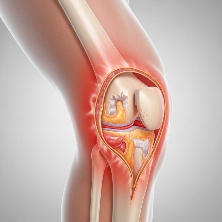
How Aponeurosis Functions
The primary function of an aponeurosis mirrors that of a tendon in many ways, but with distinct mechanical advantages. Aponeuroses attach your muscles to your bones, enabling the transmission of force generated by muscle contractions to the skeletal system. When you contract your muscles by flexing or extending a joint, the aponeurosis functions similarly to a spring, enduring the tension and pressure created by muscular contraction. This elastic quality is essential for smooth, coordinated movement.
Beyond force transmission, aponeuroses serve several additional functions within your body:
Energy Absorption and Distribution
When your muscles contract and generate force, aponeuroses absorb the energy created by this activity. Their sheet-like structure allows them to distribute this energy across a larger surface area, reducing stress concentration on any single point. This energy absorption capability helps protect your skeletal system from excessive impact and contributes to the efficiency of movement patterns. The elastic properties of aponeurotic tissue enable it to store and release energy in a manner similar to springs, which can enhance athletic performance and reduce fatigue.
Muscle Coverage and Protection
Aponeuroses envelop muscles, providing a protective layer that helps maintain muscle organization and integrity. This coverage creates distinct compartments within your body where muscles are grouped functionally and anatomically. The protective layer formed by aponeuroses helps prevent muscles from shifting during movement and contributes to the structural organization of different body regions.
Force Distribution
The broad, flat surface of an aponeurosis allows muscular forces to be distributed across a wider area of bone or cartilage rather than concentrated at a single point. This distribution mechanism reduces the risk of localized stress injuries and helps ensure that forces are transmitted more evenly throughout the skeletal system. This is particularly important in areas where muscles have broad origins or insertions.
Aponeurosis vs. Tendons: Understanding the Differences
While aponeuroses and tendons share similar functions and composition, several key differences distinguish these structures:
| Feature | Aponeurosis | Tendon |
|---|---|---|
| Shape | Flat, sheet-like structure | Thick, cord-like structure |
| Appearance | Broad, translucent sheets | Cylindrical or cordlike formations |
| Force Distribution | Spreads force across broader areas | Concentrates force transmission |
| Coverage Area | Covers large muscle regions | Connects individual muscles to bones |
| Flexibility | More spread-out tension distribution | More localized force transmission |
| Body Locations | Abdominal wall, palm of hand, foot | Achilles tendon, biceps tendon |
Both structures are composed of dense connective tissue rich in collagen fibers, but their organizational patterns and functional applications differ. Aponeuroses are particularly common in areas where large muscle groups need coordinated action, while tendons are found where individual muscles require attachment to specific skeletal points.
Location of Aponeurosis in Your Body
Aponeuroses are distributed throughout your body in various anatomically significant locations:
Abdominal Wall
The anterior abdominal wall contains several important aponeuroses, including those formed by the external oblique, internal oblique, and transversus abdominis muscles. These aponeuroses interweave to form the rectus sheath, which encloses the rectus abdominis muscle. This complex arrangement of aponeurotic tissue provides structural support for your abdominal organs and contributes to core stability. The posterior lamella of the internal oblique aponeurosis plays a particularly important role in abdominal wall reconstruction procedures, as it can be surgically modified to enhance fascial advancement and tension management.
Hand and Foot
The palmar aponeurosis in the hand and the plantar aponeurosis in the foot are prominent sheet-like structures that provide support and stability to these structures. The plantar aponeurosis, also called the plantar fascia, helps maintain the arch of your foot and distributes forces during walking and running. The palmar aponeurosis provides similar structural support and force distribution in the hand.
Head and Neck
The scalp contains the epicranial aponeurosis (also called the galea aponeurotica), which connects the frontal and occipital muscles. This structure helps coordinate facial expressions and provides structural support to the scalp.
Back and Shoulder
The thoracolumbar fascia is an extensive aponeurotic structure in the back that provides support for spinal muscles and contributes to trunk stability and movement coordination.
Clinical Significance
Understanding aponeurosis is important for healthcare professionals and patients alike, as these structures play important roles in various clinical scenarios. Injuries to aponeurotic tissue, whether from acute trauma or chronic strain, can affect movement and function. Conditions such as plantar fasciitis demonstrate how aponeurotic tissue can become inflamed or damaged, leading to pain and functional limitations. The structure of aponeuroses also has important implications for surgical procedures, particularly in abdominal wall reconstruction, where understanding fascial tension and advancement is crucial for successful outcomes.
Surgical interventions involving aponeuroses require careful technical consideration, as these structures bear significant mechanical loads. The quantitative assessment of tension on fascial elements during surgical procedures has become increasingly important for optimizing outcomes and minimizing complications. Healthcare providers must consider how various myofascial releases affect both anterior and posterior fascial elements to ensure appropriate tension reduction without excessive tissue manipulation.
Common Aponeurosis-Related Conditions
Several conditions can affect aponeurotic tissue and impact your movement and comfort:
Plantar Fasciitis
This condition involves inflammation of the plantar aponeurosis in the foot, causing heel pain and affecting walking. The condition often results from repetitive strain or excessive tension on the aponeurotic tissue.
Abdominal Wall Hernias
Weakness or separation of the aponeuroses in the abdominal wall can lead to hernias, where abdominal contents protrude through the compromised tissue. Surgical repair often involves reconstruction of these aponeurotic layers to restore structural integrity.
Dupuytren’s Contracture
This condition involves abnormal thickening and contraction of the palmar aponeurosis, causing the fingers to curl and limiting hand function.
Injury and Recovery
Aponeurotic injuries can range from minor strains to more significant tears. The recovery process depends on the severity of the injury, the location of the damage, and individual healing capacity. Minor aponeurotic injuries often respond well to conservative treatment, including rest, ice, compression, and elevation (RICE protocol). More severe injuries or conditions may require physical therapy, anti-inflammatory medications, or surgical intervention.
The vascular supply to aponeurotic tissue is relatively limited compared to muscle tissue, which can somewhat slow the healing process. However, the organized structure of aponeurotic tissue often allows for effective healing and restoration of function when appropriate treatment is provided.
Frequently Asked Questions
Q: What is the main difference between an aponeurosis and a tendon?
A: The primary difference lies in their structure and shape. Aponeuroses are broad, flat sheets of connective tissue that distribute force across larger areas, while tendons are thick, cord-like structures that concentrate force transmission at specific points. Both attach muscles to bones but function in different anatomical contexts.
Q: Where can aponeuroses be found in the body?
A: Aponeuroses are found throughout your body in areas requiring broad muscle attachments and force distribution. Common locations include the abdominal wall, palm of the hand, sole of the foot, scalp, back, and shoulder regions.
Q: What is the function of aponeurosis?
A: Aponeuroses serve multiple functions: they attach muscles to bones to enable movement, absorb and distribute energy from muscle contractions, provide protective coverage for muscles, and distribute forces across broader skeletal areas to reduce stress concentration.
Q: Can aponeurosis be injured?
A: Yes, aponeurotic tissue can be injured through acute trauma or chronic strain. Plantar fasciitis and abdominal wall hernias are common conditions involving aponeurotic structures. Most minor injuries respond to conservative treatment, while more severe cases may require professional medical intervention.
Q: How does aponeurotic tissue heal?
A: Aponeurotic tissue heals through the inflammatory and remodeling phases typical of connective tissue healing. Although aponeuroses have limited blood supply compared to muscle, they generally heal effectively with appropriate rest, conservative treatment, and sometimes physical therapy.
Q: Why is understanding aponeurosis important for surgeons?
A: Understanding aponeurotic structure and function is crucial for surgical procedures, particularly in abdominal wall reconstruction. Surgeons must carefully assess fascial tension and plan myofascial releases to optimize outcomes and minimize complications such as neurovascular injuries.
References
- Aponeurosis — Cleveland Clinic. 2022-07-05. https://my.clevelandclinic.org/health/body/23407-aponeurosis
- Aponeurosis | Fibrous Tissue, Connective Tissue, & Muscles — Britannica. https://www.britannica.com/science/aponeurosis
- Quantitative Tension on the Abdominal Wall in Posterior Component Separation with Transversus Abdominis Release — JAMA Surgery, Cleveland Clinic Center for Abdominal Core Health. 2023. https://jamanetwork.com/journals/jamasurgery/fullarticle/2810111
- Quantitative Tension on the Abdominal Wall in Posterior Component Separation — National Center for Biotechnology Information (NCBI). 2023. https://pmc.ncbi.nlm.nih.gov/articles/PMC10551814/
Similar Articles


Read full bio of Sneha Tete