Asymmetrical Pigmented Follicular Openings
Essential dermoscopic guide to asymmetrical pigmented follicular openings in lentigo maligna diagnosis and differentiation.

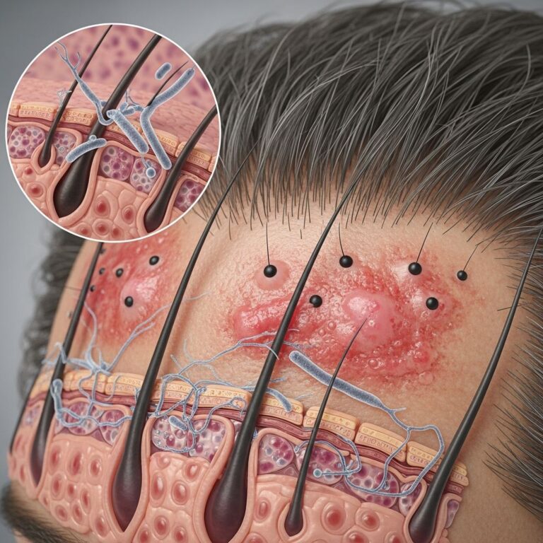
Asymmetrical pigmented follicular openings represent a critical dermoscopic feature primarily associated with lentigo maligna (LM), also known as lentigo maligna melanoma in situ. This structure appears as uneven pigmentation around hair follicle openings, often crescent-shaped or irregular, aiding in the non-invasive diagnosis of facial melanomas.
What are asymmetrical pigmented follicular openings?
Asymmetrical pigmented follicular openings (APFO) are dermoscopic structures characterized by irregular, often crescentic or partial ring-like pigmentation encircling hair follicle ostia unevenly. Unlike symmetric pigmentation in benign lesions, APFO shows one-sided or patchy hyperpigmentation, typically darker and grayish, reflecting atypical melanocyte infiltration down follicles.
Histopathologically, APFO correlates with uneven proliferation of single atypical melanocytes or small nests extending asymmetrically along the follicular epithelium into the basal layer. This nonuniform descent creates the crescent-shaped appearance visible en face under dermoscopy.
Clinical significance
APFO is a highly specific indicator of LM, with sensitivity around 47-81% and specificity up to 96% when combined with other features like dark rhomboidal structures and slate-gray dots/globules. Early identification via dermoscopy enables precise biopsy site selection, crucial for sun-damaged facial skin where margins are irregular.
In progression models, APFO emerges as the initial dermoscopic sign of LM, preceding short lines, rhomboidal structures, and eventual follicular obliteration in invasive lentigo maligna melanoma (LMM).
Dermoscopic patterns of hyperpigmented follicular openings
Hyperpigmented follicular openings in LM exhibit five distinct patterns, ranging from subtle to overt asymmetry:
- Fine circle: Thin, uniform ring around follicle, mildly asymmetric.
- Semicircle: Crescent-shaped pigmentation on one side (classic APFO).
- Signet ring-like circle: Thickened, eccentric ring resembling a signet ring.
- Irregular circle: Distorted, jagged ring with variable thickness.
- Double circle (circle within a circle/isobar): Concentric rings with darker central dot; highly specific for LM (4.2% sensitivity) but rare in actinic keratosis.
In LM, pigmentation is typically grayish-brown and darker than surrounding areas, unlike the matching tone in solar lentigos.
Histopathological correlation
Reflectance confocal microscopy (RCM) and histopathology confirm APFO as nonuniform atypical melanocyte infiltration along follicular circumference. Single units or nests descend follicles unevenly, producing crescentic pigmentation.
As lesions progress:
- Initial: Asymmetric single melanocytes in basal epidermis/follicles.
- Intermediate: Confluent nests form lines/rhomboids around adnexa.
- Advanced: Dermal melanophages create gray dots/granular patterns; follicles obliterate.
Differential diagnosis
APFO must be differentiated from benign mimics on photoaged facial skin. Key distinctions:
| Feature/Lesion | Lentigo Maligna (LM) | Solar Lentigo | Pigmented Actinic Keratosis (PAK) |
|---|---|---|---|
| Pigmentation Symmetry | Asymmetric crescents | Symmetric, matching background | May be asymmetric but regular dots |
| Color/Hue | Grayish-brown, darker | Homogeneous brown | Orange-reddish, slate-gray dots |
| Associated Patterns | Rhomboids, gray globules | Regular network | Annular-granular, absent asymmetry |
| Specificity for Malignancy | High (81% in LM) | Benign | Low; biopsy if asymmetric |
Solar lentigos show symmetric hyperpigmented follicles blending with background. PAK may mimic with asymmetry but lacks pronounced crescents; regular slate-gray dots favor PAK. Scalp LM exhibits similar APFO alongside irregular pseudonetworks.
Related dermoscopic structures
Dots aggregated around adnexal openings
Brown dots (junctional melanocyte aggregates) and blue-gray granules (dermal melanophages) cluster perifollicularly, evolving from APFO.
Short and polygonal lines
Coalesced dots form zig-zag lines around/between follicles, progressing to rhomboidal structures—polygonal extensions indicating confluent nests.
Pigmented rhomboidal structures
Thickened polygonal shapes from merged lines; highly specific for LM (combined sensitivity 89%, specificity 96%).
Annular-granular pattern
Perifollicular gray dots/granules; correlates with melanophages. Symmetric in benign, asymmetric/clustered in LM.
Obliteration of follicular openings
Advanced LM/LMM: Follicles filled/erased by pigment, often with scar-like/whitish areas.
Diagnostic algorithms
For facial pigmented lesions:
- Identify pseudonetwork (facial-specific).
- Scan for APFO (crescents/signet rings).
- Check for rhomboids/gray dots (confirm LM).
- Biopsy if ≥2 malignant features; monitor symmetric patterns.
Multivariate models: APFO + rhomboids + slate-gray dots/globules = 89% sensitivity, 96% specificity. Scalp lesions require caution due to overlap with actinic keratosis/solar lentigo.
Extragacial and scalp presentations
Though classic on face, APFO occurs in extrafacial LM (e.g., scalp) with irregular pigmentation and follicular invasion. No anterior-posterior scalp differences noted. Three cases showed rhomboids + asymmetric follicles.
Management implications
Dermoscopy-guided biopsy targets APFO areas, improving margin control in staged excision. RCM enhances recurrence detection (100% sensitivity vs. 47% dermoscopy).
Frequently Asked Questions (FAQs)
Are asymmetrical pigmented follicular openings pathognomonic for lentigo maligna?
No, while highly specific (81%), they can rarely appear in pigmented actinic keratosis; biopsy confirms.
How does APFO differ from solar lentigo follicles?
LM: Asymmetric crescents, grayish hue; solar lentigo: Symmetric, same color as background.
What is the ‘circle within a circle’ sign?
Double concentric pigmentation (isobar); specific for LM (4.2% sensitivity).
Can APFO predict LM progression to invasive melanoma?
Yes, precedes rhomboids, granular patterns, and follicular obliteration in LMM.
Is dermoscopy reliable for scalp LM diagnosis?
Moderate; overlaps with benign lesions—use flowcharts for accuracy.
References
- Lentigo Maligna – dermoscopedia — Dermoscopedia. 2023. https://dermoscopedia.org/Lentigo_Maligna
- Dermoscopy of pigmented lesions of the face: a diagnostic challenge — BVSALUD. 2022. https://fi-admin.bvsalud.org/document/view/z2x8j
- Lentigo Maligna: Keys to Dermoscopic Diagnosis — Actas Dermo-Sifiliográficas. 2016-10-01. https://actasdermo.org/en-lentigo-maligna-keys-dermoscopic-diagnosis-articulo-S1578219016300877
- Dermoscopy of Lentiginous Melanomas and Equivocal Benign … – NIH — PubMed Central (NIH). 2024-01-29. https://pmc.ncbi.nlm.nih.gov/articles/PMC10866176/
- Grey circles on dermoscopy – DermNet — DermNet NZ. 2023. https://dermnetnz.org/topics/grey-circles-dermoscopy
Similar Articles


Read full bio of medha deb