Atopy Patch Test: Diagnosis and Clinical Applications
Understanding atopy patch testing for diagnosing delayed hypersensitivity in atopic dermatitis.

Atopy Patch Test: Definition and Overview
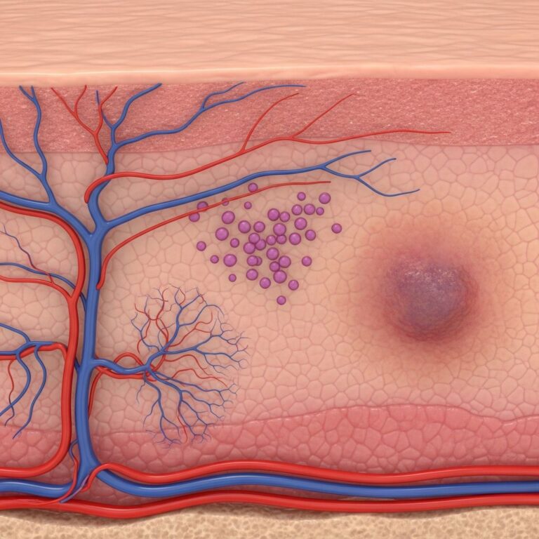
The atopy patch test (APT) is a specialized diagnostic procedure used to assess delayed-type hypersensitivity reactions to protein allergens known to provoke immunoglobulin E (IgE)-mediated responses in atopic individuals. Unlike conventional patch testing, which evaluates contact allergens through hapten-mediated reactions, the atopy patch test employs intact protein allergens applied to the skin surface for a defined period, typically 48 hours, to evaluate type IV (delayed) hypersensitivity reactions. The test represents an important advancement in understanding the complex immunological mechanisms underlying atopic dermatitis and identifying clinically relevant allergen sensitization.
Atopic dermatitis is increasingly understood as a chronic inflammatory condition involving both IgE-mediated and non-IgE-mediated immune dysregulation. The skin barrier function is compromised, likely due to filaggrin mutations, which allows enhanced entry of environmental allergens and irritants. This impaired barrier function, combined with abnormal immune responses, creates a susceptibility to allergen sensitization that can exacerbate disease manifestations.
Immunological Basis and Hypersensitivity Types
Two primary types of hypersensitivity reactions are implicated in atopic dermatitis:
- Type I IgE-mediated hypersensitivity: Immediate reactions diagnosed through skin prick tests and measurement of serum-specific immunoglobulin E levels
- Type IV T cell-mediated hypersensitivity: Delayed reactions assessed using patch testing methodologies, including the specialized atopy patch test for protein allergens
The atopy patch test bridges a critical diagnostic gap by identifying type IV sensitization to aeroallergens and food allergens in patients who may be negative on type I testing but still demonstrate clinical symptoms consistent with allergen exposure. This capability is particularly valuable in patients with multiple IgE sensitizations where clinical relevance remains unclear, or in those with severe, persistent atopic dermatitis where triggering factors have not been identified through conventional diagnostic approaches.
Differences Between Atopy Patch Test and Standard Patch Test
While both procedures involve epicutaneous application of test substances, important distinctions exist between atopy patch testing and conventional contact sensitization patch testing:
| Feature | Atopy Patch Test | Standard Patch Test |
|---|---|---|
| Allergen Type | Intact protein allergens | Haptens and chemical allergens |
| Hypersensitivity Type | Type IV in atopic individuals with IgE sensitization | Type IV contact sensitization |
| Test Duration | 48 hours | 48 hours (evaluated at 96 hours) |
| Clinical Application | Atopic dermatitis, food/aeroallergen evaluation | Contact dermatitis diagnosis |
| Reading Methodology | ETFAD standardized reading key (erythema and papule assessment) | Standard dermatitis reaction scoring |
Indications for Atopy Patch Testing
The atopy patch test is particularly valuable in several clinical scenarios:
- Suspected aeroallergen or food allergen sensitization: Patients with clinical history suggesting allergen involvement but negative specific IgE or skin prick test results
- Severe or persistent atopic dermatitis: Cases where disease remains difficult to control despite conventional management and trigger factors remain unidentified
- Multiple IgE sensitizations with unclear relevance: Patients demonstrating positive IgE testing to multiple allergens but unclear clinical correlation with symptoms
- Food allergen assessment: Evaluation of potential food triggers in patients with gastrointestinal and dermatological symptoms suggestive of food sensitivity
- Aeroallergen-pattern correlation: Cases where distribution of eczematous lesions suggests airborne allergen exposure
Contraindications and Safety Considerations
Certain clinical situations warrant against performing atopy patch testing:
- Acute exacerbations of atopic dermatitis, particularly on areas designated for testing
- Pregnancy and lactation
- Immunodeficiency disorders
- Ongoing immunosuppressive pharmacotherapy
- Autoimmune diseases
- Compromised skin integrity with active infection or severe inflammation at test sites
Test Materials and Allergen Preparation
Optimal atopy patch test results depend critically on allergen quality and preparation. Purified allergens with known protein content demonstrate superior diagnostic accuracy compared to crude extracts. Allergen concentrations are standardized using protein nitrogen units (PNU) for aeroallergens, with optimal concentrations ranging from 5 to 7 PNU, and 1% weight by volume for food allergens. Notably, allergen concentrations required for atopy patch testing exceed those used in skin prick testing, reflecting the different immunological pathways being evaluated.
The European Task Force on Atopic Dermatitis (ETFAD) has established standardized protocols specifying that intact protein allergens should be purified in petrolatum base. These allergen-petrolatum mixtures are applied to filter paper loaded into 12-millimeter aluminum test cups mounted on Scanpor tape, ensuring consistent contact between allergen and skin during the test period.
Procedural Technique and Patient Preparation
The atopy patch test procedure follows a standardized protocol to maximize reliability and reproducibility. Allergen extracts, prepared using a dropper, are placed on individual filter papers inserted into 12-mm aluminum test cups. These cups are then affixed to the upper back using medical-grade adhesive tape, typically on non-abraded, non-irritated skin. Patient compliance is essential, and individuals are counseled to avoid activities that could dislodge the test chambers, including vigorous exercise, excessive sweating, and prolonged water exposure.
The test chambers remain in place for 48 hours, at which point the first clinical evaluation occurs. A second reading is performed at 72 hours, allowing assessment of delayed hypersensitivity reactions that may develop progressively. The ETFAD reading key is employed for standardized evaluation, assessing the degree of erythema (redness) and the number, morphology, and distribution pattern of papules (small raised lesions) at each test site.
Clinical Interpretation and Results
Positive atopy patch test reactions manifest as localized dermatitis at the application site, appearing as erythema (redness), edema (swelling), and papulation (small raised bumps). Vesiculation and blistering may occur in intensely positive reactions. The morphological characteristics and distribution pattern of reactions, as assessed by the ETFAD reading system, provide clinically meaningful information about the degree and type of sensitization.
A significant association has been documented between positive atopy patch test results for aeroallergens and an air-exposed distribution pattern of eczematous lesions. Notably, strong atopy patch test positivity to house dust mites correlates with more extensive atopic dermatitis, suggesting that sensitization to this ubiquitous aeroallergen plays a substantial role in determining disease severity. This relationship underscores the potential clinical utility of the test in identifying major contributory factors to disease perpetuation.
Clinical Significance and Food Allergen Assessment
The clinical relevance of atopy patch test results is particularly evident in food allergen evaluation. Patients demonstrating positive atopy patch test reactions to specific food allergens frequently experience relief from atopic flares and associated gastrointestinal complaints upon dietary elimination of the offending food. This direct correlation between test positivity and clinical improvement validates the test’s utility in identifying clinically meaningful food sensitivities.
Comparative studies have revealed interesting geographic and population-specific variations in allergen significance. In an Indian study population, food allergens were found to be less significant contributors to atopic dermatitis compared to aeroallergens, suggesting that environmental and genetic factors influence the relative importance of different allergen categories. Additionally, the atopy patch test has been employed to identify unexpected allergen triggers, such as specific food additives, with carmine (a red food coloring derived from insects) being implicated in atopic dermatitis evolution in certain patients.
Predictive Value and Diagnostic Accuracy
The predictive accuracy of atopy patch testing has been evaluated in comparison with other diagnostic modalities. In studies predicting oral food challenge outcomes, the atopy patch test demonstrated 100% negative predictive value, indicating excellent sensitivity for excluding clinically irrelevant sensitization. However, positive predictive values vary across different tests, with skin prick testing showing the highest positive predictive value (0.91), followed by radioallergosorbent testing (0.82), and atopy patch testing (0.63). These findings suggest that while atopy patch testing is valuable for excluding sensitization, additional clinical correlation and potentially oral food challenges may be necessary to confirm clinically relevant food allergy in some patients.
Research Applications and Pathomechanism Elucidation
Beyond clinical diagnosis, the atopy patch test serves as a valuable research tool for investigating the immunopathogenic mechanisms of atopic dermatitis. Positive atopy patch test reactions correlate with positive lymphocyte transformation tests and the presence of allergen-specific T helper 2 (Th2) cells in peripheral blood, establishing a connection between cutaneous test reactivity and systemic immune response patterns. This relationship enables researchers to investigate how allergen exposure perpetuates the chronic inflammatory state characteristic of atopic dermatitis and how avoidance of identified allergens may interrupt disease pathogenesis.
Limitations and Clinical Considerations
Despite its diagnostic utility, atopy patch testing has several limitations affecting its widespread adoption. The test requires expensive, standardized allergen preparations that may have limited commercial availability. Reimbursement for atopy patch testing varies considerably among healthcare systems, creating barriers to routine use. Additionally, as even minor variations in test procedure influence sensitivity, specificity, and reproducibility, the test requires careful standardization and operator expertise to ensure reliable results.
The test may reveal type IV sensitization in patients who are completely negative for corresponding type I (IgE-mediated) tests, addressing a significant diagnostic gap in conventional allergy testing. However, the clinical relevance of type IV sensitization to aeroallergens and food allergens requires individual assessment in the context of patient history and symptom patterns.
Frequently Asked Questions
Q: How does the atopy patch test differ from a skin prick test?
A: The skin prick test evaluates immediate, IgE-mediated hypersensitivity reactions appearing within 15-20 minutes, while the atopy patch test assesses delayed, type IV hypersensitivity reactions that develop over 48-72 hours. The atopy patch test uses intact protein allergens applied to larger skin surface areas, whereas skin prick tests involve intradermal injection of small allergen amounts.
Q: Can atopy patch testing predict the outcome of oral food challenges?
A: Atopy patch testing has demonstrated utility in predicting oral food challenge results, with excellent negative predictive value (100% in some studies), meaning a negative test effectively excludes clinically relevant food allergy. However, positive predictive values are more modest, indicating that positive results require clinical correlation and may necessitate confirmatory testing.
Q: What should I expect during the atopy patch test procedure?
A: The procedure involves application of allergen-containing patches to your upper back, which remain in place for 48 hours. You must avoid strenuous exercise, excessive sweating, and activities that might dislodge the patches. The first reading occurs at 48 hours after application, with a final assessment at 72 hours. Positive reactions appear as localized redness, swelling, or small raised bumps at the application site.
Q: Is the atopy patch test suitable for all patients with atopic dermatitis?
A: No, the test is contraindicated in patients experiencing acute eczema flares, those who are pregnant or breastfeeding, individuals with immunodeficiency or autoimmune diseases, and those receiving immunosuppressive medications. Testing should be performed on non-irritated, intact skin for optimal results.
Q: What clinical benefits result from identifying positive atopy patch test allergens?
A: Identifying positive allergens enables targeted avoidance strategies, which can significantly reduce atopic flares and associated symptoms including gastrointestinal complaints in food-allergic individuals. Aeroallergen identification may guide environmental modifications or targeted immunological interventions to improve disease control.
References
- Atopy patch test — Indian Journal of Dermatology, Venereology and Leprology. 2024. https://ijdvl.com/atopy-patch-test/
- The atopy patch test for food and inhalants — PubMed, National Center for Biotechnology Information. 2013. https://pubmed.ncbi.nlm.nih.gov/23857067/
- Patch Testing for Skin Allergies — Nationwide Children’s Hospital Family Resources. 2025. https://www.nationwidechildrens.org/family-resources-education/health-wellness-and-safety-resources/helping-hands/patch-testing-for-skin-allergies
- Patch Testing for Skin Allergies: Procedure & Results — Cleveland Clinic Health Information. 2025. https://my.clevelandclinic.org/health/diagnostics/patch-test
- What You Should Know About Patch Testing and Contact Dermatitis — Tufts Medicine. 2024. https://www.tuftsmedicine.org/about-us/news/what-you-should-know-about-patch-testing-and-contact-dermatitis
- Patch testing can find what’s causing your rash — American Academy of Dermatology. 2024. https://www.aad.org/public/diseases/eczema/types/contact-dermatitis/patch-testing-rash
Similar Articles


Read full bio of Sneha Tete