Atypical Melanocytic Naevus: Clinical Features and Management
Understanding atypical melanocytic naevi: diagnosis, risk assessment, and clinical management strategies.

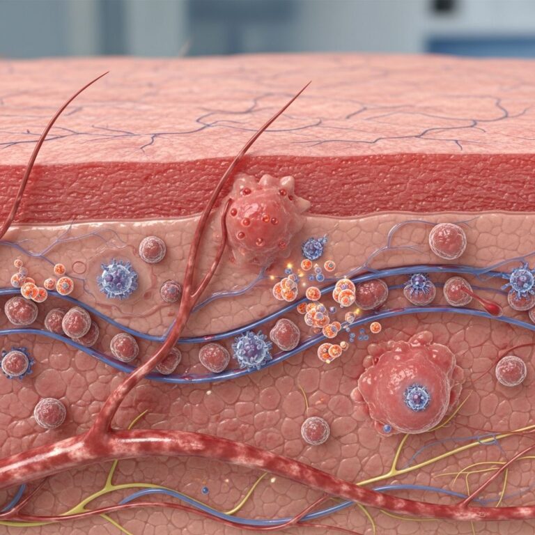
Atypical melanocytic naevi (AMN) are skin lesions that occupy a unique position on the proliferative continuum between benign melanocytic naevi and melanoma. These lesions are characterized by distinctive clinical and histological features that distinguish them from typical benign moles while requiring careful differentiation from malignant melanoma. Understanding their presentation, diagnosis, and management is essential for clinicians and patients at increased risk.
Definition and Classification
Atypical melanocytic naevi, also known as dysplastic naevi, Clark naevi, or B-K moles, are acquired melanocytic neoplasms with disordered architecture and atypia of melanocytes. The term dysplastic naevus specifically refers to the histological appearance of these lesions, which demonstrate abnormal morphological changes under microscopic examination.
These lesions are fundamentally benign, meaning they are not cancerous at the time of diagnosis. However, their clinical significance lies in their association with increased melanoma risk in affected individuals. The distinction between the clinical diagnosis of atypical moles and the histological diagnosis of dysplastic naevi is important to recognize, as only a minority of clinically atypical moles meet strict histopathological dysplastic criteria.
Atypical Mole Syndrome
Atypical Mole Syndrome (AMS) represents a distinct clinical entity defined by the presence of multiple atypical naevi in an individual. The formal diagnostic criteria for AMS include an individual with more than 50 naevi, of which three or more demonstrate atypical features.
AMS can occur in two forms:
- Sporadic dysplastic nevus syndrome: Development of multiple atypical moles without a family history of melanoma
- Familial dysplastic nevus syndrome (FAMMM): Multiple atypical moles with a significant family history of melanoma, often inherited in an autosomal dominant pattern
In cases where patients develop more than 100 lesions with a diameter greater than 8 mm, a diagnosis of dysplastic nevus syndrome is particularly warranted and requires more intensive surveillance protocols.
Clinical Presentation and Morphological Features
Atypical melanocytic naevi present with distinctive clinical characteristics that may overlap with features of melanoma. The clinical diagnosis is generally based on the presence of three or more of the following features:
- Diameter larger than 5-6 mm (often compared to the size of a pencil eraser)
- Poorly defined or ill-defined borders
- Irregular or scalloped margins
- Color variation within the lesion, typically displaying shades of tan, brown, and black
These lesions often demonstrate what clinicians describe as a ”fried-egg” appearance, characterized by a central raised or papular area surrounded by a flat, irregularly pigmented halo.
ABCDE Criteria for Evaluation
Because atypical moles clinically resemble melanoma, they are commonly evaluated using the established ABCDE criteria for melanoma detection:
| Criterion | Description |
|---|---|
| A – Asymmetry | One half of the lesion is visibly different from the other half |
| B – Border | Irregular, scalloped, or poorly defined borders rather than smooth, distinct edges |
| C – Color | Varied coloration with multiple shades of tan, brown, black, or sometimes white, red, or blue |
| D – Diameter | Lesions typically greater than 6 mm at diagnosis, though smaller lesions may be concerning |
| E – Evolving | Moles that appear different from others or show changes in size, shape, or color |
Any mole displaying one or more of these features warrants examination by a dermatologist. Additionally, other changes such as lesions that begin to burn, itch, bleed, or display surface changes should be reported and evaluated.
Clinical Subtypes and Variations
Atypical melanocytic naevi exhibit phenotypic variability that can include distinct subtypes:
- Targeted subtype: Features a central annular zone with variable pigmentation, resembling a target-like appearance
- Erythematous subtype: Displays characteristic pink pigmentation with few or sparse pigmentation remnants
Individual moles vary considerably in appearance, and some atypical moles may be larger than surrounding naevi, while others may show prominent color heterogeneity or notably jagged borders. This variability reflects the underlying architectural disorder characteristic of these lesions.
Location and Age of Onset
Atypical moles typically begin to appear at or around puberty and continue to develop into early adulthood. While they can occur anywhere on the body, they most commonly develop on the trunk, including the chest, back, and abdomen. Facial involvement is relatively rare, though atypical moles may occasionally develop on the scalp, head, and neck areas.
The development of atypical moles in elderly patients over the age of 60 is uncommon and is often regarded as an early sign of melanoma in situ or nevoid lentigo maligna, warranting careful evaluation and possible biopsy.
Histopathological Features
Microscopically, atypical melanocytic naevi demonstrate distinctive features that define the dysplastic appearance. These include architectural disorder with lentiginous or junctional component proliferation, variable cell density, and cytological atypia of melanocytes. The presence and degree of these features determine whether a lesion meets histopathological criteria for dysplastic naevus.
The histological evaluation is crucial because it helps establish the level of atypia present and provides additional information regarding melanoma risk. However, it is important to note that histological dysplasia alone does not predict individual lesion progression to melanoma.
Melanoma Risk Assessment
Risk stratification is a critical component of atypical mole management. While individual atypical naevi infrequently progress to melanoma, the presence of multiple atypical moles significantly increases melanoma risk at the patient level.
Patients with increased melanoma risk include those with:
- Multiple atypical moles (often more than 50 total naevi)
- A personal history of melanoma
- First-degree relatives (parent, sibling, or child) with melanoma
- Second-degree relatives (grandparent, grandchild, aunt, or uncle) with melanoma
- Features consistent with FAMMM syndrome
Conversely, individuals with only a few atypical moles (one to four) and no family history or personal history of melanoma have a small increased risk of developing melanoma compared to the general population.
Clinical Diagnosis and Biopsy Recommendations
The diagnosis of atypical melanocytic naevi is generally made clinically based on morphological assessment. However, because distinction between atypical moles and melanoma based on macroscopic examination alone can be difficult, many lesions are biopsied by healthcare providers to exclude malignancy.
Dermatologists often employ dermoscopy as a useful diagnostic tool to differentiate atypical moles from superficial spreading melanoma. However, when clinical concern or diagnostic uncertainty exists, lesions should be managed following melanoma treatment guidelines, typically through excisional or punch biopsy.
A biopsy involves removing the mole or a representative portion for microscopic examination, allowing pathological confirmation of the diagnosis and assessment of dysplasia grade.
Differential Diagnosis
The key differential diagnosis for atypical melanocytic naevi is melanoma, particularly the superficial spreading subtype. Other lesions that may be confused with atypical moles include:
- Congenital naevi: Usually present with larger diameter and prominent hairs, which are absent in acquired atypical moles
- Seborrheic keratosis: Characterized by a dull surface appearance and hyperkeratosis, facilitating clinical differentiation in most cases
- Lentigo and lentigo maligna: Typically present in sun-exposed areas with different morphological characteristics
Management and Surveillance Protocols
Management of atypical melanocytic naevi depends on the clinical context and individual risk factors. An atypical mole does not automatically require treatment simply because of its atypical appearance. Instead, management focuses on surveillance and early detection of melanoma.
Recommended surveillance protocols include:
- Regular full-body skin examinations: Every 3-6 months for individuals with AMS or FAMMM syndrome, beginning at puberty. Examination frequency may be reduced once moles appear stable and mature
- Baseline skin photography: Whole-body and targeted photographs to document mole appearance and track changes over time
- Patient self-examination: Monthly self-assessment using the ABCDE criteria to identify changes in existing moles or development of new concerning lesions
- Family screening: Recommendation that first-degree and second-degree relatives undergo dermatological evaluation, as dysplastic nevus syndrome often shows familial aggregation
Biopsy or removal may be indicated when a specific lesion exhibits suspicious features suggesting possible melanoma, when changes are observed during surveillance, or when a lesion becomes symptomatic (bleeding, itching, or changing in appearance).
Additional Screening Considerations
Dermatologists may recommend supplemental screening measures for high-risk patients:
- Ophthalmological examination: Evaluation by an ophthalmologist, as dysplastic nevus syndrome may be associated with ocular findings in some familial cases
- Genetic counseling: For families meeting FAMMM criteria, genetic evaluation and testing may be appropriate
- Advanced imaging: Reflectance confocal microscopy or other advanced dermatological imaging techniques may assist in differentiating atypical moles from melanoma in selected cases
Prognostic Considerations
It is important to emphasize that atypical melanocytic naevi themselves have a low rate of malignant transformation. The increased melanoma risk associated with these lesions reflects an overall patient phenotype rather than a high probability that any individual atypical mole will become cancerous.
However, the cumulative risk of melanoma development increases significantly with the total number of atypical moles present. Larger numbers of atypical and typical melanocytic naevi correlate with greater melanoma risk. When detected and properly treated in early stages, melanoma has a high cure rate, making surveillance and early detection strategies crucial for improved outcomes.
Clinical Pearls and Key Takeaways
Several important clinical points guide the evaluation and management of atypical melanocytic naevi:
- Atypical moles are benign lesions that require careful monitoring but not routine removal
- The ABCDE criteria provide a systematic approach to identifying concerning lesions
- Multiple atypical moles (AMS) warrant more intensive surveillance than isolated atypical moles
- Family history of melanoma significantly impacts risk stratification and surveillance intensity
- Regular dermatological evaluation and patient education are cornerstone interventions
- Histopathological examination clarifies diagnosis but does not reliably predict individual lesion progression
Frequently Asked Questions
Q: Are atypical moles cancerous?
A: No, atypical moles are benign lesions. However, individuals with multiple atypical moles have an increased risk of developing melanoma compared to those without atypical moles. This risk reflects overall skin phenotype rather than malignant transformation of individual moles.
Q: How often should I have my moles checked if I have atypical moles?
A: If you have Atypical Mole Syndrome (more than 50 naevi with three or more atypical), dermatological screening every 3-6 months is recommended. Individuals with fewer atypical moles may require less frequent evaluation. Your dermatologist will determine appropriate intervals based on your specific risk factors.
Q: Do all atypical moles need to be removed?
A: No. Atypical moles are removed only when there is clinical suspicion for melanoma, when a lesion shows concerning changes during surveillance, or when a patient experiences symptoms like itching or bleeding. Routine removal of stable atypical moles is not recommended.
Q: Can atypical moles appear on the face?
A: Atypical moles rarely appear on the face, but they may develop on the scalp, head, and neck regions. The trunk (chest, back, abdomen) is the most common location for atypical mole development.
Q: What should I do if a mole starts changing?
A: Any change in a mole’s size, shape, color, or symptoms (itching, bleeding, tenderness) warrants evaluation by a dermatologist. These changes may indicate melanoma or another concerning process requiring prompt professional assessment.
Q: Is there a genetic component to atypical mole syndrome?
A: Yes. While sporadic dysplastic nevus syndrome can occur without family history, familial dysplastic nevus syndrome (FAMMM) shows autosomal dominant inheritance patterns. If you have a family member with melanoma or atypical moles, your risk may be elevated, and genetic counseling may be beneficial.
References
- Atypical (Dysplastic) Melanocytic Naevus — Professional Clinical Guidance, Pediatric Dental & Medical Association. May 16, 2025. https://www.pcds.org.uk/clinical-guidance/atypical-dysplastic-melanocytic-naevus
- Atypical Nevus or Atypical Mole — Water’s Edge Dermatology. https://www.wederm.com/service/atypical-nevus/
- Atypical Mole — StatPearls, National Center for Biotechnology Information (NCBI). https://www.ncbi.nlm.nih.gov/books/NBK560606/
- Dysplastic Nevi — StatPearls, National Center for Biotechnology Information (NCBI). https://www.ncbi.nlm.nih.gov/books/NBK482210/
- Atypical Moles: Diagnosis and Management — American Family Physician. June 1, 2015. https://www.aafp.org/pubs/afp/issues/2015/0601/p762.html
- Atypical Nevi (Moles) — Merck Manuals Professional Edition. https://www.merckmanuals.com/professional/dermatologic-disorders/benign-skin-tumors-growths-and-vascular-lesions/atypical-nevi-moles
Similar Articles


Read full bio of medha deb