Atypical Mycobacterial Infection: 4 Syndromes & Treatments
Comprehensive guide to infections caused by non-tuberculous mycobacteria, including causes, symptoms, diagnosis, and treatment options.

Atypical mycobacterial infections are caused by species of mycobacterium other than Mycobacterium tuberculosis, the agent of tuberculosis, and Mycobacterium leprae, the cause of leprosy. These non-tuberculous mycobacteria (NTM), also known as atypical mycobacteria, are ubiquitous in the environment, including water, soil, and animals, leading to opportunistic infections particularly in immunocompromised individuals or following trauma.
What are the clinical features of atypical mycobacterial infection?
Atypical mycobacterial infections manifest in four main clinical syndromes: skin and soft tissue infections, lymphadenitis, disseminated disease, and bone and joint infections. Skin and soft tissue infections are the most common extrapulmonary form encountered by dermatologists, often presenting with nonspecific lesions such as papules, nodules, plaques, abscesses, ulcers, or sporotrichoid patterns (linear distribution along lymphatics).
Rapidly growing mycobacteria like M. fortuitum, M. chelonae, and M. abscessus typically cause localized skin lesions post-trauma, surgery, or cosmetic procedures, presenting as erythema, edema, abscesses, or non-healing ulcers. Slow-growing species such as M. marinum (fish tank granuloma) produce nodules along inoculation sites, while M. ulcerans (Buruli ulcer) leads to painless necrotic ulcers, predominantly on extremities.
Lymphadenitis is common in children, especially with M. avium complex (MAC), presenting as unilateral cervical swelling. Disseminated infections occur in severely immunocompromised patients (e.g., HIV/AIDS), with multiple skin lesions like erythematous papules or nodules, often alongside systemic symptoms. Bone and joint infections involve deeper tissues, causing osteomyelitis or septic arthritis.
Incubation periods vary: weeks for rapid growers to months for slow growers. Lesions may mimic sporotrichosis, tuberculosis, nocardiosis, or fungal infections, complicating diagnosis. In immunosuppressed patients, suppuration predominates, while immunocompetent hosts show granulomatous reactions.
How is atypical mycobacterial infection diagnosed?
Diagnosis requires a high index of suspicion, especially for persistent lesions unresponsive to standard antibiotics. History of trauma, surgery, water exposure, or immunosuppression is key.
Clinical examination reveals heterogeneous lesions without pathognomonic features, except for classic entities like fish tank granuloma (sporotrichoid nodules post-aquarium injury) or Buruli ulcer (necrotic ulcer).
- Swabs or biopsies: From ulcers, pus, or tissue for culture on Lowenstein-Jensen medium or automated systems like MGIT.
- Histopathology: Shows suppurative granulomas, mixed inflammation, or neutrophilic abscesses; acid-fast bacilli (AFB) staining (Ziehl-Neelsen) has low sensitivity (11-90%, higher in immunocompromised).
- Culture and speciation: Gold standard, but slow (weeks to months); rapid growers culture faster.
- Molecular tests: PCR for speciation and resistance profiling, though less available for cutaneous cases.
- Imaging: Radiographs or MRI for deep infections.
Differentials include cutaneous TB, leprosy, fungal infections, leishmaniasis, and pyoderma gangrenosum.
What is the treatment for atypical mycobacterial infection?
Treatment is organism-specific, guided by culture and sensitivity, often requiring prolonged combination antibiotics due to resistance. Surgical intervention is frequently needed.
| Organism Group | Common Antibiotics | Duration | Surgical Role |
|---|---|---|---|
| Rapid Growers (M. fortuitum, chelonae, abscessus) | Amikacin + Clarithromycin; Ciprofloxacin + TMP-SMX; Imipenem | 4-6 months | Aggressive debridement essential |
| M. marinum | Clarithromycin + Ethambutol or Rifampicin + TMP-SMX | 3-6 months | Debridement if needed |
| M. ulcerans (Buruli) | Rifampicin + Clarithromycin | 8 weeks | Wide excision ± grafting |
| MAC (disseminated) | Clarithromycin + Ethambutol + Rifabutin | 12+ months | Lymph node excision |
General regimen includes rifampicin, ethambutol, isoniazid, minocycline, ciprofloxacin, clarithromycin, azithromycin, or cotrimoxazole. Monitor for side effects like hepatotoxicity. Some lesions heal spontaneously with scarring, especially in immunocompetent hosts.
In pediatrics, lymphadenitis may resolve spontaneously or require excision. Postsurgical infections demand thorough debridement.
What is the outcome for atypical mycobacterial infection?
Prognosis varies: excellent for localized skin infections with prompt treatment (resolution in months), but poor for disseminated disease in AIDS patients without immune reconstitution. Scarring, lymphedema, or contractures occur post-Buruli ulcer. Relapse is common if treatment is inadequate; resistance is rising.
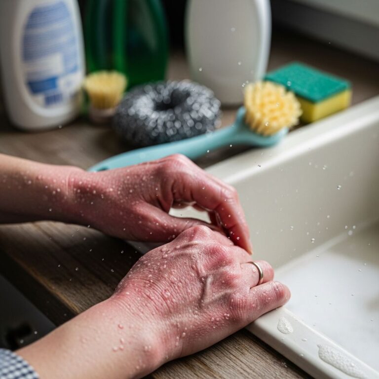
Prevention: Avoid contaminated water post-surgery, use sterile techniques, and consider prophylaxis in high-risk groups.
Frequently Asked Questions
Who is at risk for atypical mycobacterial infections?
Immunocompromised individuals (HIV, transplant recipients), post-surgical patients, children (lymphadenitis), and those exposed to contaminated water or soil.
Can atypical mycobacterial infections spread person-to-person?
No, they are not contagious; acquired environmentally.
Do all lesions require antibiotics?
No, some self-resolve, but treatment prevents complications; culture guides therapy.
How long does treatment last?
Typically 3-12 months, depending on organism and response.
Is surgery always needed?
Often for abscesses, nodes, or necrotic lesions, combined with antibiotics.
Related Topics
- Cutaneous tuberculosis
- Leprosy
- Sporotrichosis
- Buruli ulcer
- Fish tank granuloma
References
- Cutaneous Atypical Mycobacterial Infections: A Brief Review — Indian Journal of Dermatology, PMC. 2024. https://pmc.ncbi.nlm.nih.gov/articles/PMC11616912/
- Mycobacterial Skin Infections — Patient.info. 2024. https://patient.info/doctor/dermatology/mycobacterial-skin-infections
- Cutaneous Atypical Mycobacterial Infections: A Brief Review — PubMed. 2024. https://pubmed.ncbi.nlm.nih.gov/39640455/
- Atypical mycobacterial infection — DermNet NZ. 2024. https://dermnetnz.org/topics/atypical-mycobacterial-infection
- Cutaneous non-tuberculous mycobacterial infections: An update — Journal of Skin and Sexually Transmitted Diseases. 2023. https://jsstd.org/cutaneous-non-tuberculous-mycobacterial-infections-an-update/
Similar Articles


Read full bio of medha deb