Atypical Solar Lentigo: Recognition and Management
Understanding atypical solar lentigines: clinical features, diagnosis, and treatment approaches.

Atypical Solar Lentigo: Clinical Recognition and Management
Solar lentigines are among the most common benign pigmented lesions encountered in dermatological practice, particularly in individuals with a history of chronic ultraviolet radiation (UVR) exposure. While most solar lentigines present with characteristic features that allow straightforward diagnosis, atypical variants can pose diagnostic challenges and require careful clinical evaluation to exclude more serious conditions such as melanoma or other malignant lesions. This comprehensive guide examines the clinical characteristics, pathophysiology, diagnostic approaches, and management strategies for atypical solar lentigines.
Definition and Clinical Features of Solar Lentigines
Solar lentigines, commonly referred to as age spots, liver spots, or sunspots, are benign melanocytic proliferations that develop in response to chronic cumulative sun exposure. These lesions are characterized by a localized increase in melanocyte activity and subsequent accumulation of melanin within keratinocytes, resulting in visible areas of hyperpigmentation. In their typical presentation, solar lentigines appear as well-demarcated, flat macules ranging from 3 to 20 millimeters in diameter. The color typically ranges from light tan to dark brown, with uniform pigmentation throughout each individual lesion.
However, atypical solar lentigines deviate from this standard presentation. When the pigmentation demonstrates an irregular shape or variable color distribution, the lesion is more accurately described as atypical. These variations may include:
- Irregular or ill-defined borders rather than clearly demarcated edges
- Heterogeneous color with multiple shades of brown, black, or gray within a single lesion
- Larger size, particularly when exceeding 10 millimeters in diameter
- Asymmetrical appearance with variation in lesion morphology
- Surface texture changes or slight elevation from the surrounding skin
The distinction between typical and atypical solar lentigines is clinically significant because atypical variants may represent either benign lesions with unusual morphological features or potentially concerning lesions that require more rigorous evaluation to exclude dysplasia or malignancy.
Pathophysiology and Mechanism of Formation
Solar lentigines are believed to represent ultraviolet radiation-induced proliferative responses of epidermal keratinocytes and/or melanocytes, although the exact mechanism of formation remains incompletely understood. Current evidence suggests that ultraviolet B (UVB) radiation increases expression of keratinocyte growth factor, which subsequently induces tyrosinase expression and melanin production in melanocytes. This melanin pigment is then transferred to keratinocytes, where abnormal pigment retention occurs, leading to the visible pigmentation characteristic of solar lentigines.
The development of atypical features may reflect variations in the degree or pattern of melanocyte proliferation, differences in melanin distribution within the epidermis, or concomitant changes in other epidermal structures. Understanding these pathophysiological mechanisms is essential for both diagnosis and treatment selection.
Epidemiology and Risk Factors
Solar lentigines predominantly affect middle-aged and older individuals, with prevalence increasing significantly after age 40. Several demographic and environmental factors substantially increase the risk of developing these lesions:
| Risk Factor | Impact on Development |
|---|---|
| Fair skin and sun sensitivity | Individuals who burn easily and do not tan efficiently are at substantially higher risk |
| Prolonged sun exposure | Sun damage accumulates over time; patients with lentigines typically have extensive cumulative UVR exposure |
| Age over 50 years | Most common presentation occurs after age 50, though fair-skinned individuals may develop lesions as early as age 30 |
| Geographic location | Higher incidence in populations living in high-UV areas |
| History of acute sunburns | Younger individuals often develop lentigines following severe sunburns |
Atypical variants appear to share similar epidemiological patterns but may show different distributions or frequencies in specific populations.
Variants of Solar Lentigines
Several distinct variants of solar lentigines merit recognition, as they may present with atypical or unusual features:
PUVA-Induced Lentigo
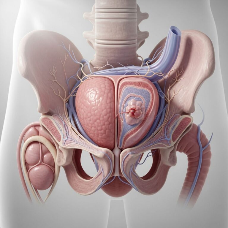
Psoralen ultraviolet A (PUVA) therapy can induce a characteristic variant of lentigo, developing in approximately 50% of patients who have received at least 6 years of PUVA treatment. These lesions may sometimes be distinguished by large, somewhat atypical melanocytes on histopathological examination. PUVA-induced lentigines can develop on any body surface exposed to PUVA therapy, including the genitalia, making them potentially concerning due to their unusual locations.
Ink Spot Lentigo
The ink spot lentigo represents another atypical variant, characterized by a distinctive appearance. These lesions appear as small black or dark gray macules resembling ink spots, with reticulated (net-like) borders. They frequently occur on the shoulders and other sun-exposed areas. The distinctive morphology with reticulated borders distinguishes them from typical solar lentigines and may cause concern regarding their nature.
Diagnostic Evaluation and Differential Diagnosis
Proper diagnosis of atypical solar lentigines requires a systematic clinical approach combining history, examination, and diagnostic techniques. The primary challenge lies in differentiating atypical solar lentigines from other pigmented lesions, particularly melanoma or melanoma in situ.
Clinical History
A thorough history should document:
- Duration of the lesion and whether it has changed over time
- History and extent of sun exposure or UV therapy
- Family history of skin cancer or atypical moles
- Any symptoms such as itching, bleeding, or discomfort
- Previous skin lesions and their outcomes
Dermatoscopic Examination
Dermatoscopic examination represents a crucial diagnostic tool for evaluating pigmented lesions. A dermatoscopic examination can help distinguish atypical solar lentigines from other conditions. Dermoscopy allows visualization of subsurface pigmentation patterns, which may reveal features consistent with benign solar lentigines versus concerning features suggestive of dysplasia or malignancy. Key dermatoscopic features of typical solar lentigines include homogeneous pigmentation and regular, parallel patterns, whereas atypical variants may show heterogeneous patterns or features requiring closer scrutiny.
Differential Diagnoses
Several conditions must be distinguished from atypical solar lentigines:
- Melanoma and melanoma in situ—characterized by asymmetry, irregular borders, color variation, and potential for growth
- Atypical nevus (dysplastic nevus)—may show features overlapping with atypical lentigines but typically have dermatoscopic features of nests or specific architectural patterns
- Melasma—symmetric patches of hyperpigmentation, often on the face, with distinct demarcation patterns differing from typical lentigines
- Seborrheic keratosis—benign proliferation that may develop atypical features or arise within pre-existing lentigines
- Lentigo maligna—a form of melanoma in situ that may present with features overlapping atypical solar lentigo
Secondary Changes and Complications
Solar lentigines may undergo secondary changes that further complicate their appearance and potentially warrant intervention. Understanding these transformations is essential for proper management.
Seborrheic Keratosis Development
Solar lentigines can sometimes develop seborrheic keratoses within the original lentigo, resulting in localized thickening and change in texture. These secondary lesions are benign but alter the morphology of the original lentigo, creating a more complex appearance. In other cases, inflammation may occur within the lesion.
Lichenoid Keratosis
A significant secondary change involves inflammation of the solar lentigo, leading to what is termed a lichenoid keratosis or lichen-planus-like keratosis. These inflamed variants exhibit a specific pattern of inflammation visible on histopathological examination. Importantly, lichenoid keratoses often resolve gradually over time without requiring intervention, distinguishing them from the persistent nature of typical solar lentigines.
Management and Treatment Options
While solar lentigines are benign and generally do not pose medical risks, patients may seek treatment for cosmetic reasons or if they become concerned about the lesion’s appearance. It is important to note that treatment is not medically necessary.
Observational Management
Many solar lentigines remain asymptomatic and unchanged over time, and patients may opt for observation without active treatment. However, lesions that change in size, color, or appearance warrant evaluation by a dermatology professional to exclude malignant transformation.
Cryotherapy (Freezing)
Cryotherapy, involving the application of liquid nitrogen to freeze the lesion, represents a popular treatment option for isolated or smaller solar lentigines. This quick, in-office procedure results in the treated area flaking off within days. One study found cryotherapy was more effective than 40% trichloroacetic acid (TCA) for treating solar lentigines, although the difference was not statistically significant. However, cryotherapy and other destructive treatments may occasionally leave temporary or permanent dark or light marks.
Laser Therapy
Laser treatments represent highly effective options for removing solar lentigines with precision. Q-switched lasers or fractional lasers target melanin in the pigmented areas to break it down with minimal damage to surrounding skin. Aggressive therapy using quality-switched lasers is effective for treating solar lentigines but carries the risk of post-inflammatory hyperpigmentation (PIH), particularly in darker skin types. For darker skin types, less intensive irradiation reduces this risk while maintaining treatment efficacy.
Intense Pulsed Light (IPL)
Intense pulsed light therapy represents another option for treating solar lentigines, offering an alternative to traditional laser therapy.
Chemical Peels
Chemical peels using topical solutions such as alpha hydroxy acids or trichloroacetic acid (TCA) exfoliate the outermost layer of skin, helping fade solar lentigines and improve overall skin tone over time.
Topical Treatments
Various topical agents have been investigated for treating hyperpigmentation. Cysteamine cream can reduce excess pigmentation over time. However, hydroquinone and other traditional bleaching creams have shown limited success in treating solar lentigines.
Treatment Considerations
Several important factors should guide treatment decisions:
- Risk of post-treatment discoloration: Most destructive treatments may leave temporary or permanent white or dark marks
- Lesion size and location: Treatment approach varies depending on these factors
- Skin type: Darker skin types require modified laser parameters to minimize PIH risk
- Patient expectations: Clear communication about potential outcomes and limitations
- Lesion characteristics: Atypical features may require biopsy before treatment rather than destructive procedures
Prevention and Sun Protection
As with many skin conditions induced by UV exposure, prevention remains the most effective approach. Regular and early use of broad-spectrum sunscreens, along with strategies to minimize sun exposure—particularly during peak UV hours—can substantially reduce the development of solar lentigines over time. Educating individuals about sun protection from a young age plays a crucial role in preventing these and other signs of photoaging.
Frequently Asked Questions
Q: How do I distinguish atypical solar lentigines from melanoma?
A: While some features overlap, atypical solar lentigines typically show irregular color or borders that are still relatively uniform, whereas melanoma usually demonstrates greater asymmetry, more irregular borders, color variation (multiple browns, blacks, or reds), and often grows over time. Dermatoscopic examination and biopsy may be necessary for definitive diagnosis.
Q: Are atypical solar lentigines dangerous?
A: Most atypical solar lentigines are benign and do not pose medical risks. However, because they may occasionally mimic more serious conditions, proper evaluation by a dermatology professional is essential, particularly when lesions exhibit atypical features.
Q: Can solar lentigines transform into cancer?
A: While benign solar lentigines themselves do not transform into melanoma, they serve as markers of chronic sun damage. Individuals with extensive solar lentigines have increased cumulative sun exposure and thus elevated overall skin cancer risk.
Q: Is treatment necessary for atypical solar lentigines?
A: Treatment is not medically necessary, as atypical solar lentigines are benign. However, patients may choose treatment for cosmetic reasons or if concerned about the lesion’s appearance. Treatment decisions should be individualized based on patient preferences and lesion characteristics.
Q: What is the most effective treatment for atypical solar lentigines?
A: Laser therapy, particularly Q-switched or fractional lasers, offers highly effective treatment with precision and minimal damage to surrounding skin. However, cryotherapy is also effective and may be preferred for isolated lesions. Treatment choice depends on lesion characteristics, skin type, and patient preferences.
Q: Will atypical solar lentigines return after treatment?
A: If treated lesions are adequately destroyed, they should not recur. However, continued sun exposure may lead to development of new solar lentigines in other areas.
References
- Solar lentigo – VisualDx — VisualDx. Accessed 2026-01-28. https://www.visualdx.com/
- Understanding Solar Lentigo: Causes and Treatment Options — Devonshire Dermatology. 2025-06-08. https://www.devonshiredermatology.co.uk/
- Solar Lentigo (Age Spots) – Causes, Symptoms & Treatment — St. Johns Physician Partners. Accessed 2026-01-28. https://www.sjpp.org/
- Lentigo (Age Spots) — Yuma Dermatology. Accessed 2026-01-28. https://www.yumadermatology.com/
- Lentigo: Causes, Symptoms, and Treatment — Patient.info. Accessed 2026-01-28. https://patient.info/
- Solar lentigo: causes and treatment options — Mesoestetic. Accessed 2026-01-28. https://www.mesoestetic.com/
- Solar lentigo — Dr. Breslavets | CMSD. Accessed 2026-01-28. https://cmsderm.ca/
Similar Articles


Read full bio of medha deb