Barrett’s Esophagus: Essential Guide To Symptoms And Treatment
Understand Barrett's esophagus: causes, symptoms, diagnosis, treatment, and cancer risk from GERD complication.

Barrett’s esophagus is a premalignant condition where the normal squamous lining of the distal esophagus is replaced by columnar epithelium, often due to chronic gastroesophageal reflux disease (GERD). This change increases the risk of esophageal adenocarcinoma, though the annual progression rate remains low at 0.2% to 0.5%.
What Is Barrett’s Esophagus?
Barrett’s esophagus occurs when the flat pink squamous cells lining the esophagus transform into thick red columnar cells resembling intestinal tissue, typically triggered by repeated acid exposure from GERD. Defined as salmon-colored mucosa extending at least 1 cm above the gastroesophageal junction with intestinal metaplasia confirmed by biopsy, it affects the distal esophagus where it meets the stomach.
The lower esophageal sphincter (LES), a valve between the esophagus and stomach, weakens in GERD, allowing stomach acid and bile to reflux upward, damaging esophageal tissue. Over time, this chronic injury prompts metaplastic change as squamous cells adapt to the acidic environment by becoming acid-resistant columnar cells with goblet cells.
Not all GERD patients develop Barrett’s esophagus; risk factors include male sex, age over 50, White race, central obesity, smoking, and family history. Approximately half of diagnosed individuals have minimal reflux symptoms, known as silent reflux.
Symptoms of Barrett’s Esophagus
Most people with Barrett’s esophagus experience GERD symptoms, including frequent heartburn—a retrosternal burning sensation worsening after meals—and acid regurgitation. Other signs include dysphagia (difficulty swallowing), chest pain, chronic cough, hoarseness, sore throat, or weight loss, though many remain asymptomatic.
- Frequent heartburn and regurgitation of stomach contents
- Difficulty or painful swallowing (dysphagia)
- Chest pain unrelated to heart disease
- Less common: nausea, vomiting, or sensation of fullness while eating
Symptoms alone do not diagnose Barrett’s; persistent GERD warrants endoscopic evaluation, especially in high-risk groups.
Causes and Risk Factors
Chronic GERD is the primary cause, where repeated acid and bile reflux inflames the esophagus, leading to cellular adaptation. Bile acids exacerbate damage by upregulating inflammatory cytokines like IL-8 and IL-1b, recruiting immune cells and promoting metaplasia.
Key risk factors include:
- Long-standing GERD (most common)
- Male gender (4-5 times higher risk than females)
- Age >50 years
- White ethnicity
- Obesity, particularly central adiposity
- Current or former smoking
- Hiatal hernia
- Family history of Barrett’s or esophageal cancer
The pathogenesis involves molecular changes: acid exposure induces inflammation, while bile promotes proliferation of stem cells at the squamocolumnar junction, leading to intestinal metaplasia.
Diagnosis
Diagnosis requires upper endoscopy (esophagogastroduodenoscopy or EGD) revealing salmon-colored mucosa ≥1 cm above the gastroesophageal junction, confirmed by biopsy showing intestinal metaplasia with goblet cells.
The Prague classification grades extent: C (circumferential length) and M (maximum length) of Barrett’s segment, aiding risk stratification. High-definition endoscopy, narrow-band imaging, and biopsies via Seattle protocol (four-quadrant every 1-2 cm) enhance detection of dysplasia.
Histopathology distinguishes:
- Nondysplastic: Columnar epithelium with goblet cells, nuclear enlargement in basal glands.
- Low-grade dysplasia (LGD): Nuclear enlargement, stratification, increased N:C ratio, mitoses in crypts.
- High-grade dysplasia (HGD): Marked atypia, complex architecture, frequent atypical mitoses.
Patients with chronic GERD symptoms >5 years, especially with alarm features (dysphagia, weight loss), should undergo screening endoscopy.
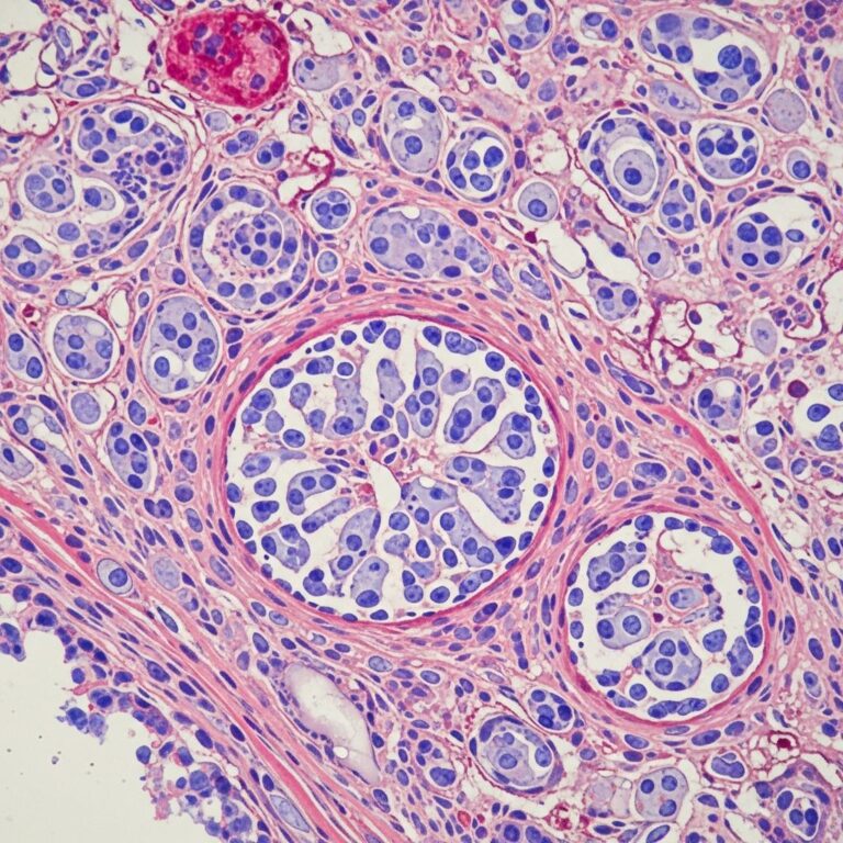
Pathophysiology and Histopathology
Chronic GERD inflammation replaces squamous epithelium with columnar mucosa resistant to acid. Progression: metaplasia → low-grade dysplasia → high-grade dysplasia → adenocarcinoma.
| Stage | Key Features | Risk |
|---|---|---|
| Metaplasia (Nondysplastic) | Goblet cells, salmon mucosa | 0.2-0.5% annual to cancer |
| Low-Grade Dysplasia | Nuclear crowding, basal stratification | ~1% annual progression |
| High-Grade Dysplasia | Surface maturation loss, lamina propria invasion | High; treat aggressively |
Image description: Endoscopic view shows tongue-like projections of red columnar mucosa replacing pale squamous epithelium proximal to gastroesophageal junction.
Complications
The primary concern is esophageal adenocarcinoma, with lifetime risk 5-10% in Barrett’s patients versus <1% in general population. Annual incidence: 0.2-0.5%; higher with dysplasia (LGD: 0.5-1%, HGD: 6-19%).
Other issues: strictures from uncontrolled reflux, ulcers, or bleeding.
Treatment
Treatment focuses on GERD control with proton pump inhibitors (PPIs) twice daily to heal esophagitis and reduce progression risk. Lifestyle: weight loss, elevate head of bed, avoid triggers (alcohol, caffeine, large meals).
Surveillance: Endoscopy intervals based on dysplasia:
- No dysplasia: Every 3-5 years
- LGD: Every 6-12 months (confirm with expert pathology)
- HGD/early cancer: Endoscopic eradication therapy (EET)
Endoscopic Therapies: For dysplasia/intramucosal cancer, radiofrequency ablation (RFA) achieves complete eradication of intestinal metaplasia (CEIM) in 80-90%, with low recurrence. Endoscopic mucosal resection (EMR) for nodular areas, followed by ablation.
Surgery (esophagectomy) reserved for invasive cancer. Emerging: potassium-competitive acid blockers (P-CABs) for refractory cases, though untested specifically.
Prevention
Prevent progression by treating GERD aggressively: high-dose PPIs, antireflux surgery (fundoplication) if needed. Smoking cessation, weight management reduce risk. Screen high-risk GERD patients; aspirin/NSAIDs may lower risk but not routinely recommended.
When to See a Doctor
Seek care for persistent heartburn > twice weekly, new dysphagia, unexplained weight loss, or vomiting. Those with long-term GERD should discuss screening.
Frequently Asked Questions (FAQs)
What causes Barrett’s esophagus?
Chronic GERD from LES dysfunction allows acid reflux, damaging and altering esophageal cells.
Does Barrett’s esophagus always lead to cancer?
No; annual risk is low (0.2-0.5%), but surveillance detects early changes.
How is Barrett’s esophagus diagnosed?
By endoscopy and biopsy confirming metaplasia ≥1 cm above GE junction.
Can Barrett’s esophagus be cured?
Dysplasia can be eradicated via ablation; nondysplastic managed with surveillance and PPIs.
Who should be screened for Barrett’s?
Men >50 with chronic GERD and risk factors (obesity, smoking).
References
- Barrett Esophagus – StatPearls — NCBI Bookshelf / StatPearls Publishing. 2023-07-17. https://www.ncbi.nlm.nih.gov/books/NBK430979/
- Barrett’s esophagus – Symptoms and causes — Mayo Clinic. 2023. https://www.mayoclinic.org/diseases-conditions/barretts-esophagus/symptoms-causes/syc-20352841
- Barrett Esophagus: A Review — JAMA Network. 2021-05-18. https://jamanetwork.com/journals/jama/fullarticle/2795263
- Barrett’s Esophagus: Symptoms and Treatment — Banner Health. 2023. https://www.bannerhealth.com/services/gastroenterology/treatment/barretts-esophagus
- Barrett’s oesophagus — Better Health Channel (Victoria.gov.au). 2023. https://www.betterhealth.vic.gov.au/health/conditionsandtreatments/barretts-oesophagus
Similar Articles


Read full bio of medha deb