Bioengineered Skin: 5 Clinical Uses And Key Benefits
Innovative skin substitutes for burns, ulcers, and wounds: advancing regeneration and healing.

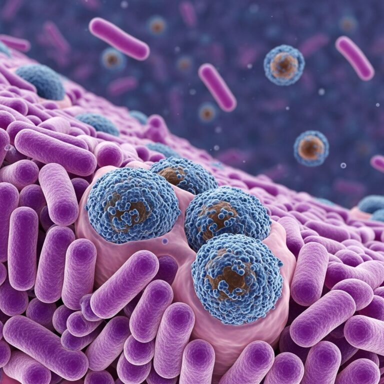
Bioengineered skin refers to laboratory-created skin substitutes designed to temporarily or permanently replace damaged or missing skin, primarily functioning to restore the epidermal barrier and support dermal regeneration until the patient’s own skin heals. These products are particularly vital for patients with extensive burns, chronic ulcers, or large wounds where traditional skin grafting is insufficient due to limited donor sites.
What is bioengineered skin?
Bioengineered skin products are developed using tissue engineering techniques, combining cells, scaffolds, and bioactive molecules to mimic the structure and function of native human skin, which consists of the epidermis, dermis, and hypodermis. They serve as temporary coverage to prevent infection, reduce fluid loss, and promote healing, or as permanent grafts that integrate with host tissue.
These substitutes address limitations of autografts (patient’s own skin) and allografts (donor skin), such as donor site morbidity and immune rejection. Early developments focused on epidermal equivalents, evolving to full-thickness dermal-epidermal constructs.
History
The concept of bioengineered skin emerged in the 1970s-1980s to meet the urgent need for skin coverage in severe burn victims. Pioneering work by Howard Green led to the first cultured epithelial autografts (CEA) in 1979, using keratinocytes expanded from small biopsies.
In the 1990s, commercial products like Epicel® (CEA) and Apligraf® (bilayered skin equivalent) gained FDA approval. Advances in the 2000s introduced dermal matrices like Integra® and AlloDerm®. Recent decades have seen integration of 3D bioprinting and stem cells for more complex, vascularized substitutes.
Types of bioengineered skin
Bioengineered skin is classified by composition and layers replicated:
- Epidermal substitutes: Consist of cultured keratinocytes forming a sheet-like epidermis. Example: Epicel® – autologous keratinocytes cultured for 2-3 weeks.
- Dermal substitutes: Provide a collagen-based matrix for fibroblast infiltration and neovascularization. Examples: Integra® (collagen-chondroitin-6-sulfate with silicone epidermis), AlloDerm® (acellular human dermis).
- Composite (dermal-epidermal) substitutes: Bilayered products with dermis and epidermis. Examples: Apligraf® (human fibroblasts in bovine collagen gel topped with keratinocytes), Orcel®.
- Full-thickness substitutes: Include subcutaneous elements, often using 3D bioprinting for hypodermis mimicry.
Temporary biological dressings like Biobrane® (silicone-nylon with porcine collagen) offer short-term coverage.
Production methods
Production involves:
- Cell harvesting: Keratinocytes and fibroblasts from skin biopsies, expanded in culture.
- Scaffolds: Biodegradable matrices like collagen, fibrin, hyaluronic acid, or synthetic polymers (e.g., polyglycolic acid).
- Cell seeding: Fibroblasts embedded in dermal scaffold, keratinocytes layered on top to form stratified epidermis.
- Bioreactor culture: Air-liquid interface promotes differentiation; growth factors (VEGF, TGF-β, PDGF) enhance vascularization.
- Quality control: Sterility, viability, absence of pathogens.
Acellular options use decellularized matrices allowing host cell repopulation.
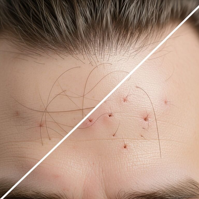
Clinical applications
Primary uses include:
- Burns: Coverage for >50% TBSA burns; CEAs reduce mortality but prone to fragility.
- Chronic wounds: Venous, diabetic, pressure ulcers unresponsive to standard care. Apligraf® accelerates closure in 4-12 weeks.
- Surgical wounds: Post-Mohs surgery defects, contractures.
- Rare diseases: Epidermolysis bullosa, modeling via humanized mice.
- Research: Disease models (psoriasis, eczema), drug testing.
| Condition | Product Example | Outcomes |
|---|---|---|
| Severe burns | Epicel® | 80-90% take rate; heals in 2-4 weeks |
| Diabetic ulcers | Apligraf® | 45% complete closure vs. 20% controls |
| Venous leg ulcers | Orcel® | Faster healing, reduced pain |
Advantages and disadvantages
Advantages
- Unlimited supply from small biopsies.
- No donor site scarring.
- Reduced infection risk, pain, blood loss.
- Improved aesthetics, elasticity vs. split-thickness grafts.
- Customizable with patient cells, genes for immune evasion (e.g., PD-L1 overexpression).
Disadvantages
- High cost ($3,000-$10,000/cm²).
- Delayed production (2-6 weeks).
- Fragility, contraction, poor pigmentation.
- Variable take rates (50-90%), shear issues.
- Immunogenicity in allogeneic products.
Effectiveness
Meta-analyses show bioengineered skin superior to standard care for chronic wounds (RR 1.95 for healing). In burns, long-term outcomes match autografts with less morbidity. Healing involves rapid re-epithelialization, angiogenesis via growth factors.
Future developments
Emerging technologies:
- 3D bioprinting: Layer-by-layer printing of cells, hydrogels for vascularized, full-thickness skin; patient-specific models for eczema, cancer.
- Stem cells: iPSCs, MSCs for self-renewal, appendages (hair, glands).
- Nanomaterials: Nanoparticles for sustained drug/growth factor release.
- Tri-layered constructs: Epidermis-dermis-hypodermis with basement membrane mimics.
- Gene editing: CRISPR for disease correction, immune tolerance.
Challenges: Vascularization, innervation, pigmentation, scalability.
Controversies and unanswered questions
- Optimal cell sources: Autologous vs. allogeneic vs. iPSC?
- Best scaffold: Natural vs. synthetic?
- Immunosuppression needs for allogeneic grafts?
- Cost-effectiveness vs. traditional methods?
- Long-term durability, cancer risk?
- Regulatory hurdles for personalized bioprinted skin?
Frequently asked questions
What is bioengineered skin used for?
Primarily for treating extensive burns, chronic non-healing wounds like diabetic foot ulcers, venous leg ulcers, and surgical defects where donor skin is limited.
How long does it take to produce bioengineered skin?
Typically 2-4 weeks for cell expansion and maturation in culture.
Is bioengineered skin permanent?
Some act as temporary dressings; others integrate permanently, supporting host regeneration over months.
Does insurance cover bioengineered skin treatments?
Often yes for FDA-approved products in indicated wounds; varies by region and insurer.
What are the success rates?
60-90% for wound closure; comparable to autografts long-term with fewer complications.
References
- Advancements in bioengineered and autologous skin grafting — Frontiers in Bioengineering and Biotechnology. 2024. https://www.frontiersin.org/journals/bioengineering-and-biotechnology/articles/10.3389/fbioe.2024.1461328/full
- Skin Bioengineering: Preclinical and Clinical Applications — Actas Dermo-Sifiliográficas. 2012. https://actasdermo.org/en-skin-bioengineering-preclinical-clinical-applications-articulo-S157821901200042X
- 3D Bioprinting: An Overview — DermNet NZ. 2024. https://dermnetnz.org/topics/3d-bioprinting
- Bioengineered skin — DermNet NZ. 2024. https://dermnetnz.org/topics/bioengineered-skin
- Skin substitutes as treatment for chronic wounds — PMC / NIH. 2023. https://pmc.ncbi.nlm.nih.gov/articles/PMC10498286/
- Challenges and promises in modeling dermatologic disorders — SAGE Journals. 2014. https://journals.sagepub.com/doi/10.1177/1535370214538747
Similar Articles


Read full bio of medha deb