Blisters And Pustules In Neonates: 4-Step Diagnostic Guide
Comprehensive guide to identifying, diagnosing, and managing blisters and pustules in newborns for parents and clinicians.

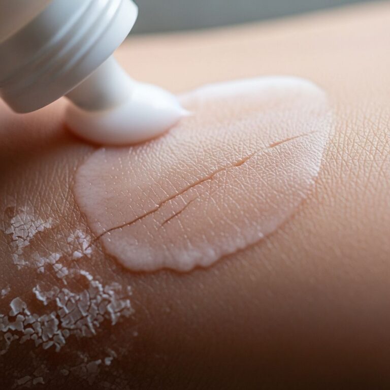
Blisters and pustules are common skin findings in newborns, often causing concern for parents and clinicians. While most are benign and self-resolving, some indicate serious infections or genetic disorders requiring prompt intervention. This article provides a structured approach to diagnosis and management based on clinical features and laboratory evaluation.
What are the clinical features of blisters and pustules in neonates?
Neonatal blisters are fluid-filled lesions greater than 0.5 cm in diameter, while pustules are smaller, pus-filled elevations. These can appear generalized or localized, with accompanying erythema, vesicles, or erosions. Benign conditions typically present in healthy, term infants feeding well, whereas systemic signs like fever, lethargy, or poor feeding suggest infection or inflammatory disease.
Distribution is key: acral involvement (hands, feet) points to transient neonatal pustular melanosis (TNPM) or acropustulosis, while napkin-area satellite pustules indicate candidiasis. Morphology aids differentiation—sterile pustules show eosinophils on smear, infectious ones neutrophils or organisms.
How are blisters and pustules in neonates classified?
Neonatal pustular dermatoses are categorized into benign self-limiting, infectious, inflammatory, genetic, immunologic, and miscellaneous groups.
- Benign self-limiting: Erythema toxicum neonatorum (ETN, 16-30% prevalence), TNPM, miliaria, neonatal cephalic pustulosis, acne.
- Infectious: Bacterial (impetigo, folliculitis), fungal (candidiasis, Malassezia), viral (herpes, varicella), parasitic (scabies).
- Inflammatory/Genetic: Eosinophilic pustular folliculitis, incontinentia pigmenti, epidermolysis bullosa.
- Immunologic: Hyper-IgE syndrome, IL-1 receptor antagonist deficiency.
- Miscellaneous: Neonatal lupus, Behcet disease.
Benign and self-limiting pustular eruptions
Erythema toxicum neonatorum
The most common neonatal pustulosis, affecting 16-30% of newborns, ETN presents day 1-3 with fleeting erythematous macules, papules, and pustules on trunk/extremities. Smear shows eosinophils; resolves in 1-2 weeks without treatment.
Transient neonatal pustular melanosis
TNPM occurs in 5% of African-American newborns, with fragile pustules at birth rupturing to hyperpigmented macules with collarette scaling. Smear: neutrophils. Benign, resolves by 3 months leaving post-inflammatory hyperpigmentation.
Miliaria (heat rash)
Sweat duct obstruction causes miliaria crystallina (clear vesicles, forehead) or rubra/pustulosa (red papules/pustules, trunk/neck at 10 days). Avoid overheating; resolves spontaneously.
Neonatal cephalic pustulosis and acne
Caused by Malassezia, presents as facial pustules weeks 2-3. Topical antifungals if persistent; acne resolves by 4 months.
Acropustulosis of infancy
Recurrent acral pustules in infants, more in darker skin; pruritic, resolves by 2-3 years. Topical steroids effective.
Infectious causes
Bacterial infections
Impetigo (S. aureus, streptococci) shows flaccid bullae/pustules with honey-crusted erosions, often periorificial. Bullous impetigo has Nikolsky sign. Treat with topical/systemic antibiotics. Folliculitis/periporitis staphylogenes secondary to miliaria. Congenital syphilis: generalized pustules, palmoplantar involvement.
Fungal infections
Candidiasis: Neonatal (acquired at birth): napkin-area beefy red plaques with satellites; congenital (intrauterine): generalized, acral/nail involvement, spares diaper area. KOH shows yeast/hyphae; treat with topical/systemic azoles. Malassezia pustulosis: Facial/scalp folliculitis, KOH/culture confirms.
Viral infections
Herpes simplex: clustered vesicles/pustules, systemic illness; Tzanck smear shows multinucleates. Varicella-zoster: evolves vesicle→pustule→crust; zoster immunoglobulin if maternal rash near delivery.
Parasitic: Scabies
Extensive vesicles/pustules/burrows on acra, genitalia, scalp/face. Not always pruritic; treat with permethrin/sulfur.
Inflammatory, genetic, and other serious causes
Serious pustulosis occurs in unwell/preterm neonates. Langerhans cell histiocytosis: scalp/trunk pustules, anemia, organomegaly. Eosinophilic pustular folliculitis: sterile crusted papules/pustules, peripheral eosinophilia.
Genetic: Incontinentia pigmenti (X-linked, vesicular stage), epidermolysis bullosa (blisters from trauma, dystrophic type ulcerates). Immunodeficiencies: Hyper-IgE (staphylococcal pustulosis, eosinophilia), DITRA (generalized pustules, fever). Neonatal lupus: annular plaques, heart block risk.
Diagnostic approach to neonatal blisters and pustules
Stepwise evaluation:
- History: Onset, maternal infections, PROM, devices, infant wellbeing.
- Exam: General/systemic, lesion distribution/morphology.
- Lab: Pustule smear (Gram, Giemsa, KOH, Tzanck), CBC, CRP, cultures, swabs. Well infant with eosinophils: benign. Neutrophils/organisms: infectious.
- Further tests: If systemic—blood/CSF cultures, serology, biopsy, genetics.
| Condition | Smear Findings | Distribution | Well/Ill |
|---|---|---|---|
| ETN | Eosinophils | Trunk/extremities | Well |
| TNPM | Neutrophils | Acral | Well |
| Candidiasis | Yeast/hyphae | Diaper/satellites | Well/Ill |
| Herpes | Multinucleates | Grouped | Ill |
| Impetigo | Gram+ cocci | Periorificial | Well/Ill |
Management of blisters and pustules in neonates
Benign: Reassurance, no treatment. Infectious: Targeted antimicrobials (e.g., mupirocin for impetigo, fluconazole for disseminated candida). Supportive: Emollients, non-adherent dressings for blisters. Avoid overheating. Sick infants: Hospitalize, sepsis workup.
Frequently Asked Questions (FAQs)
Q: Are neonatal pustules always dangerous?
A: No, most are benign like ETN or TNPM in healthy infants; smear confirms sterility.
Q: When should I seek medical help for newborn blisters?
A: If infant is unwell, feverish, poor feeding, or lesions widespread/acral in preterm.
Q: How to differentiate ETN from infection?
A: ETN has eosinophils on smear, no organisms, infant well; infections show pathogens, systemic signs.
Q: Does TNPM leave scars?
A: No, hyperpigmentation fades by 3 months without scarring.
Q: Can candidiasis be congenital?
A: Yes, intrauterine exposure causes generalized lesions including palms/soles, unlike postnatal diaper-limited.
References
- Pustular lesions in the neonate: Focused diagnostic approach based on clinical clues — Indian Journal of Dermatology, Venereology and Leprology. 2019. https://ijdvl.com/pustular-lesions-in-the-neonate-focused-diagnostic-approach-based-on-clinical-clues/
- Common rashes in neonates — Royal Australian College of General Practitioners (RACGP). 2012-05. https://www.racgp.org.au/afp/2012/may/common-rashes-in-neonates
- Neonatal Pustular Dermatosis: An Overview — PubMed Central (PMC). 2015. https://pmc.ncbi.nlm.nih.gov/articles/PMC4372928/
- Acropustulosis in infants: Symptoms, causes, and treatment — Medical News Today. 2023. https://www.medicalnewstoday.com/articles/325413
- Newborn Skin: Part I. Common Rashes — American Academy of Family Physicians (AAFP). 2008-01-01. https://www.aafp.org/pubs/afp/issues/2008/0101/p47.html
- Transient Neonatal Pustular Melanosis (TNPM) — Cleveland Clinic. 2023. https://my.clevelandclinic.org/health/diseases/23111-transient-neonatal-pustular-melanosis-tnpm
- Blisters and Erosions in the Neonate — American Academy of Pediatrics (AAP) NeoReviews. 2011. https://publications.aap.org/neoreviews/article/12/8/e453/91390/Blisters-and-Erosions-in-the-Neonate
Similar Articles


Read full bio of Sneha Tete