Blueberry Muffin Syndrome: Causes, Diagnosis, Treatment
Understanding the causes, diagnosis, and management of blueberry muffin syndrome in newborns.

Author: DermNet NZ Editor — Reviewed: January 2026
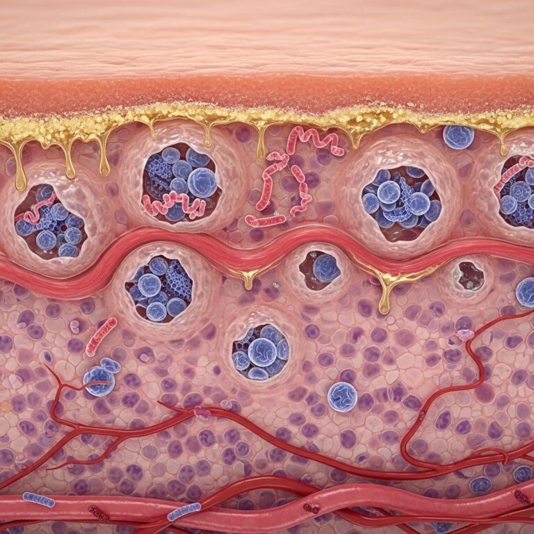
‘Blueberry muffin syndrome’ is the descriptive term used for an infant born with multiple blue/purple marks or nodules in the skin. These lesions resemble the appearance of blueberries scattered on a muffin, hence the name. The condition arises from dermal extramedullary hematopoiesis (blood cell production in the skin), purpura (bleeding into the skin), or cutaneous metastases from malignancies.
What is the cause of blueberry muffin syndrome?
During normal fetal development, hematopoiesis (blood cell formation) occurs in multiple sites, including the liver, spleen, and skin, up to the fifth month of gestation. After this period, it primarily shifts to the bone marrow. Blueberry muffin lesions in newborns indicate persistent extramedullary hematopoiesis beyond this gestational window, or other pathological processes like hemorrhage or tumor spread.
There are many underlying causes for blueberry muffin syndrome, broadly categorized into three main groups: congenital infections, hematological disorders, and neoplasms. Each category requires specific diagnostic approaches and treatments.
Congenital infections
Congenital infections, particularly those in the TORCH group (Toxoplasmosis, Other agents, Rubella, Cytomegalovirus, Herpes simplex), are classic causes. These infections trigger exaggerated extramedullary erythropoiesis as the fetus compensates for anemia or systemic stress.
- Toxoplasmosis: Caused by Toxoplasma gondii, often from undercooked meat or cat feces exposure. It leads to chorioretinitis, hydrocephalus, and intracranial calcifications alongside skin lesions.
- Rubella: Historically the most associated, coined in the 1960s during rubella outbreaks. Congenital rubella syndrome includes sensorineural deafness, cataracts, and cardiac defects.
- Cytomegalovirus (CMV): The most common congenital infection, causing microcephaly, hearing loss, and hepatosplenomegaly.
- Herpes simplex virus (HSV): Typically HSV-2, presenting with vesicles that may evolve into blueberry-like nodules.
- Other agents: Includes syphilis, parvovirus B19 (causing severe fetal anemia), coxsackievirus, varicella-zoster, hepatitis B, and Epstein-Barr virus.
Hematological disorders
Blood disorders that cause hemolytic anemia or stimulate erythropoietin production lead to dermal blood cell clusters.
- Haemolytic disease of the newborn (erythroblastosis fetalis): Due to Rh or ABO incompatibility, causing severe anemia and extramedullary hematopoiesis.
- Hereditary spherocytosis: A genetic red cell membrane defect leading to hemolysis.
- Twin-to-twin transfusion syndrome: Recipient twin develops polycythemia and hyperviscosity.
- Congenital dyserythropoietic anaemia: Rare bone marrow failure syndromes.
- Infantile pyknocytosis: Transient hemolysis in newborns.
Neoplastic disorders
Malignancies in neonates are rare but critical to exclude, as they cause firmer, larger lesions from metastatic deposits.
- Neuroblastoma: Most common, with skin metastases appearing as firm nodules.
- Congenital leukaemia: Acute myeloid or lymphoblastic leukaemia infiltrating the skin.
- Langerhans cell histiocytosis (LCH): Proliferative disorder of histiocytes.
- Rhabdomyosarcoma: Soft tissue sarcoma.
- Extraskeletal Ewing sarcoma: Rare primitive neuroectodermal tumor.
Clinical features
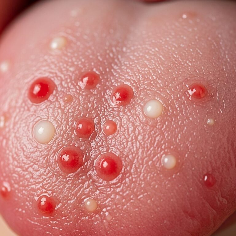
Blueberry muffin syndrome presents at birth or within the first few days of life. Lesions are 1–10 mm blue to purple macules, papules, or nodules, non-blanching, and distributed preferentially on the face, trunk, and proximal extremities. They do not weep or ulcerate. The number varies from dozens to hundreds. Associated findings depend on the cause: jaundice, hepatosplenomegaly, thrombocytopenia, or systemic illness in infections; pallor and anemia in hematological causes; firm nodules in malignancies.
The rash typically fades over 3–6 weeks, leaving hyperpigmented macules, but persistence suggests neoplasm.
Differential diagnosis
Several conditions mimic blueberry muffin syndrome:
| Condition | Key Features | Differentiator |
|---|---|---|
| Infantile haemangioma | Grows postnatally, blanchable, may ulcerate | Dynamic evolution, Doppler ultrasound |
| Cutaneous mastocytosis | Urticate on stroking (Darier’s sign) | Histology shows mast cells |
| Transient neonatal pustular melanosis | Pustules rupture to hyperpigmented macules | Sterile pustules on exam |
| Neonatal lupus erythematosus | Annular plaques on face/scalp | Maternal autoantibodies, EKG changes |
| Pyogenic granuloma | Solitary, friable, vascular | Single lesion |
Diagnosis
Diagnosis requires a systematic approach combining history (maternal infections, blood group), full physical exam, and targeted investigations.
- Blood tests: Full blood count (anemia, thrombocytopenia), blood film, Coombs test, reticulocyte count, maternal TORCH serology.
- Imaging: Cranial ultrasound (calcifications), abdominal ultrasound (hepatosplenomegaly, tumors), skeletal survey if malignancy suspected.
- Specific tests: CMV PCR, rubella IgM, toxoplasma PCR, parvovirus serology.
- Biopsy: Skin punch biopsy for histology (erythropoiesis vs. metastases), immunohistochemistry if needed.
- Other: Ophthalmological exam, hearing screen, bone marrow aspirate in persistent cases.
Management
Treatment targets the underlying cause; the rash itself is self-limited.
- Infections: Antivirals (ganciclovir for CMV), antiparasitics (pyrimethamine/sulfadiazine for toxoplasmosis), supportive care.
- Hematological: Phototherapy for jaundice, exchange transfusion for severe hemolysis, IVIG.
- Neoplastic: Chemotherapy, surgery, stem cell transplant; multidisciplinary oncology input.
- Supportive: Monitor for complications like anemia, infection; parental reassurance.
Prognosis
Prognosis varies widely by etiology. Benign causes (e.g., self-limited pyknocytosis) resolve fully with excellent outcomes. TORCH infections carry risks of neurodevelopmental delay (e.g., 50–90% hearing loss in rubella). Malignancies have guarded prognosis, with neuroblastoma stage-dependent survival rates of 70–90% for low-risk. Early diagnosis improves outcomes through prompt intervention.
Frequently Asked Questions
What does blueberry muffin syndrome look like?
It appears as numerous 1–10 mm blue-purple non-blanching macules, papules, or nodules on the trunk, face, and limbs of newborns, resembling blueberries on a muffin.
Is blueberry muffin syndrome dangerous?
It signals underlying pathology ranging from benign to life-threatening (infections, anemias, cancers), necessitating urgent evaluation.
How is blueberry muffin syndrome diagnosed?
Through clinical exam, blood tests (CBC, TORCH screen), imaging, and skin biopsy to identify the cause.
Does blueberry muffin syndrome go away?
Lesions typically fade in 3–6 weeks after treating the cause, leaving temporary hyperpigmentation.
Can blueberry muffin syndrome be prevented?
Prevent maternal infections via vaccination (rubella), hygiene, prenatal screening; Rh prophylaxis prevents hemolytic disease.
References
- Blueberry muffin baby — Wikipedia. 2023-10-01. https://en.wikipedia.org/wiki/Blueberry_muffin_baby
- Blueberry muffin syndrome — DermNet NZ. 2024-05-15. https://dermnetnz.org/topics/blueberry-muffin-syndrome
- Blueberry Muffin Rash Causes & Treatment — Cleveland Clinic. 2025-08-20. https://my.clevelandclinic.org/health/symptoms/blueberry-muffin-rash
- Blueberry muffin rash from rubella: Pictures, causes, and more — Medical News Today. 2024-11-12. https://www.medicalnewstoday.com/articles/blueberry-muffin-rash-rubella
- Practice parameter: Evaluation of the child with microcephaly — American Academy of Neurology. 2023-06-01. https://www.neurology.org/doi/10.1212/WNL.0b013e318291d96e
- Congenital CMV infection — CDC. 2025-02-14. https://www.cdc.gov/cmv/congenital-infection.html
Similar Articles


Read full bio of medha deb