Bone Scan: Complete Guide To Procedure, Uses, And Risks
Discover how bone scans detect hidden bone issues, from cancer spread to fractures, with this detailed patient guide on procedure, benefits, and safety.

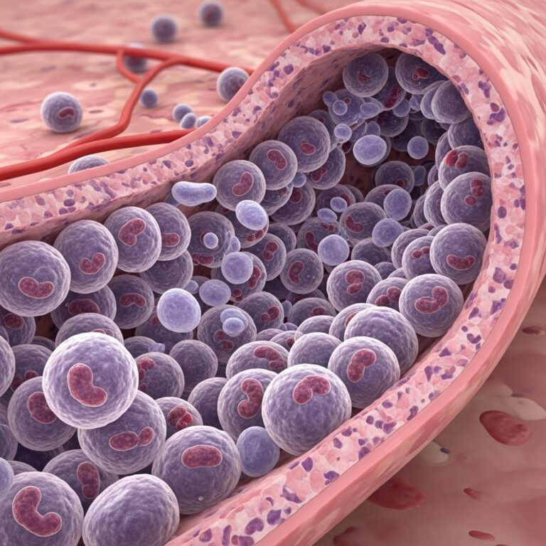
A bone scan, also known as skeletal scintigraphy, is a nuclear medicine imaging test that reveals abnormalities in bones by detecting areas of increased or decreased metabolic activity. It uses a small amount of radioactive tracer to highlight issues like fractures, infections, tumors, or metastatic cancer that standard X-rays might miss.
Understanding Bone Scans in Modern Diagnostics
Bone scans play a crucial role in evaluating skeletal health, particularly when symptoms like unexplained pain or suspected cancer spread arise. Unlike conventional radiographs, which show bone structure, this test visualizes bone turnover at a cellular level, making it highly sensitive for early detection.
The procedure involves injecting a radiotracer, typically Technetium-99m linked to a diphosphonate like MDP or HDP, which binds to areas of active bone remodeling. A gamma camera then captures emitted gamma rays to produce detailed images.
Key Medical Applications of Bone Scans
Bone scans are versatile tools ordered for various conditions. They excel in scenarios where precise localization of bone pathology is needed.
- Detecting metastatic disease: Commonly used to check if cancers from breast, prostate, lung, kidney, or thyroid have spread to bones.
- Identifying infections: Highly effective for diagnosing osteomyelitis, with sensitivity up to 94%.
- Finding hidden fractures: Reveals stress fractures in hips, feet, legs, or spine not visible on X-rays.
- Assessing metabolic bone diseases: Evaluates conditions like osteoporosis, Paget’s disease, hyperparathyroidism, or osteomalacia.
- Evaluating pain sources: Pinpoints causes of unexplained bone pain.
- Other uses: Monitors treatment response, checks bone graft viability, screens for child abuse, or assesses avascular necrosis in patients with sickle cell disease or steroid use.
In cancer care, bone scans help stage disease and plan therapies by mapping osteoblastic activity.
Step-by-Step Procedure: What Happens During a Bone Scan
The entire process is outpatient, painless, and typically takes 3-5 hours, including wait time.
- Preparation and arrival: Wear comfortable clothing; metal objects are removed. An IV line is inserted in your arm or hand.
- Tracer injection: A nuclear medicine technologist administers the radiotracer intravenously. It’s safe, with radiation less than a standard X-ray.
- Waiting period: Rest for 1-4 hours as the tracer circulates and accumulates in bones. Drink plenty of water to aid elimination; you can often leave the facility.
- Imaging phase: Lie on a table under a gamma camera that moves slowly over your body for 30-60 minutes. Remain still; multiple views may be taken. For three-phase scans (e.g., infection checks), images are captured immediately, then 3-4 hours later.
- Completion: IV is removed; resume normal activities. Tracer clears via urine within 24 hours.
Three-phase bone scans include blood flow, blood pool, and delayed phases for detailed infection or inflammation assessment.
Preparation Tips for Your Bone Scan
Minimal preparation is needed, ensuring accessibility.
- Inform staff of pregnancy, breastfeeding, allergies, recent imaging, or medications like calcium supplements.
- Hydrate well post-injection; void bladder before scanning.
- No fasting required; eat normally.
- For children or anxious patients, sedation is rare but possible.
| Patient Group | Special Considerations |
|---|---|
| Children | May need sedation; parental presence encouraged. |
| Pregnant/Breastfeeding | Discuss risks; may delay or use alternatives. |
| Recent barium exams | Wait 1-2 weeks to avoid interference. |
Safety Profile and Potential Risks
Bone scans are safe with low radiation exposure (equivalent to 1-2 years of background radiation), posing minimal risk to adults. No acute side effects; rare allergic reactions to tracer occur.
Radiation considerations: Avoid close contact with infants/young children for 24 hours post-scan. Breastfeeding may pause briefly.
Specificity can drop in complicated cases (e.g., post-surgery, hardware), requiring follow-up CT/MRI.
Interpreting Your Bone Scan Results
Images show ‘hot spots’ (increased uptake) indicating high turnover from cancer, fractures, or infection; ‘cold spots’ suggest reduced activity like early avascular necrosis.
- Normal: Even tracer distribution across bones; kidneys visible.
- Abnormal: Focal hot spots (metastases, infection); diffuse uptake (‘superscan’) in widespread disease like prostate cancer.
- Incidental findings: Soft tissue uptake may signal tumors or effusions.
Radiologist reports integrate findings with history; further tests often follow positives.
Comparing Bone Scans to Other Imaging
| Modality | Strengths | Limitations | Best For |
|---|---|---|---|
| Bone Scan | Whole-body, sensitive to early changes | Less specific; no anatomy detail | Screening metastases, infections |
| X-ray | Quick, low cost, structural view | Misses early/occult issues | Obvious fractures |
| CT | Detailed anatomy | Higher radiation, local only | Confirming bone scan findings |
| MRI | Soft tissue, no radiation | Expensive, not whole-body | Spine/foot details |
| PET | Metabolic + anatomy (with CT) | Costly | Cancer staging |
Bone scans are often first-line for broad screening due to sensitivity.
Patient Experiences and FAQs
Patients report the injection pinch as the only discomfort; lying still is key. Videos show calm procedures.
Frequently Asked Questions
Is a bone scan painful?
No, only minor IV prick; scanner is non-invasive.
How long do results take?
Preliminary same day; full report in 1-2 days.
Can I drive after?
Yes, no sedation effects.
Does it detect all cancers?
Sensitive for blastic metastases; lytic ones may need SPECT/CT.
Follow-up needed?
Often yes, for confirmation.
Advances and Future Directions
Hybrid SPECT/CT improves specificity by combining function and anatomy. New tracers enhance detection of specific pathologies. Research continues for lower radiation protocols.
References
- Bone scan – UCSF Benioff Children’s Hospitals — UCSF Benioff Children’s Hospitals. 2023. https://www.ucsfbenioffchildrens.org/medical-tests/bone-scan
- Bone Scan – StatPearls — NCBI Bookshelf. 2023-10-30. https://www.ncbi.nlm.nih.gov/books/NBK531486/
- Bone Scan — RadiologyInfo.org. 2024. https://www.radiologyinfo.org/en/info/bone-scan
- Bone Scan — UMass Memorial Health. 2023. https://www.ummhealth.org/health-library/bone-scan
- How to Prepare for a Bone Scan — American Cancer Society. 2023. https://www.cancer.org/cancer/latest-news/how-to-prepare-for-a-bone-scan.html
- What Does a Whole-Body Bone Scan Show? — Cleveland Clinic. 2023-08-01. https://my.clevelandclinic.org/health/diagnostics/17642-whole-body-bone-scan
Similar Articles


Read full bio of Sneha Tete