Botryomycosis Pathology: Clinical, Histology, Treatment Guide
Detailed histopathological analysis of botryomycosis, a rare bacterial pseudomycosis mimicking fungal infections in skin and viscera.

Botryomycosis represents a rare chronic granulomatous bacterial infection that primarily affects the skin but can involve visceral organs, characterized by the formation of distinctive bacterial granules resembling fungal structures—hence its name derived from Greek botrys (bunch of grapes) and mykosis (fungal infection). Despite its mycetoma-like appearance, it is caused by bacteria, most commonly Staphylococcus aureus, leading to a pseudomycotic reaction.
Introduction
Botryomycosis, also known as bacterial pseudomycosis or pyoderma vegetans, is an uncommon condition with approximately 200 cases reported worldwide. It manifests as a chronic suppurative and granulomatous response to bacterial infection, predominantly cutaneous (about 75% of cases) but occasionally visceral. The disease develops slowly over months to years, often in immunocompromised hosts, and features pathognomonic Splendore-Hoeppli phenomenon on histopathology.
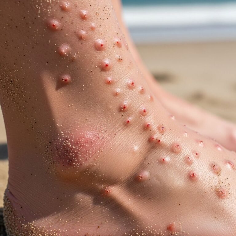
The infection typically follows local trauma, surgery, or foreign body implantation, with predisposing factors including diabetes mellitus, HIV, alcoholism, cystic fibrosis, prolonged corticosteroid use, hematologic malignancies, and other immunodeficiencies. Cutaneous lesions predominate on extremities, trunk, face, and perianal regions, presenting as nodules, abscesses, sinuses, fistulae, ulcers, and verrucous plaques with purulent discharge containing yellow grains.
Clinical Features
Cutaneous botryomycosis evolves insidiously, with lesions developing over weeks to months before ulcerating. Common presentations include:
- Subcutaneous nodules: Firm, tender masses that may suppurate.
- Abscesses and sinuses: Draining pus with characteristic yellow “grains” or granules, reminiscent of actinomycosis sulfur granules.
- Ulcerated plaques: Vegetating, verrucous surfaces with central necrosis or eschar.
- Fistulae tracts: Communicating with deeper tissues, leading to chronic drainage.
Extremities are most affected, followed by trunk and head/neck regions. In disseminated cases, multiple erythematous papules, pustules, and eschars appear across face, limbs, and mucosa, as seen in immunocompromised patients. Visceral forms, rarer, primarily involve lungs (post-surgery or in cystic fibrosis), with potential spread to liver, kidneys, brain, or bone.
Immunosuppressed patients, such as those with leukemia, may present with rapid ulceration atypical of the usual chronic course.
Histopathology
The hallmark of botryomycosis pathology is the presence of basophilic bacterial colonies (grains) surrounded by an eosinophilic radial rim known as the Splendore-Hoeppli phenomenon. This phenomenon arises from antigen-antibody complexes, fibrin, and tissue debris forming club-like projections around non-filamentous bacterial aggregates.
Microscopic Features
- Epidermis: Acanthosis, hyperkeratosis, pseudoepitheliomatous hyperplasia.
- Dermis: Dense mixed inflammatory infiltrate (neutrophils, lymphocytes, plasma cells, eosinophils); abscess formation; granulation tissue.
- Grains: 0.5-2 mm amorphous basophilic centers (bacterial colonies) with peripheral eosinophilic Splendore-Hoeppli material, radiating in “asteroid” or “sunburst” pattern.
- Deep tissues: Extension to subcutis, muscle, bone with granulomatous reaction.
Special stains enhance visualization:
| Stain | Findings |
|---|---|
| Gram stain | Gram-positive cocci clusters in grains (e.g., S. aureus) |
| Giemsa | Bacterial morphology and Splendore-Hoeppli radiating clubs |
| Periodic acid-Schiff (PAS) | Negative for fungi; highlights eosinophilic rim |
| Silver stain (Gomori methenamine) | Bacterial aggregates without fungal hyphae |
| Tissue Gram | Confirms non-acid-fast bacteria |
Unlike true mycetomas (filamentous actinomycetes/nocardia) or eumycetomas (fungal hyphae), botryomycosis grains contain coccoid bacteria without filaments.
Splendore-Hoeppli Phenomenon
This pathognomonic feature represents an immune response to bacterial antigens, forming eosinophilic sheaths around colonies. It distinguishes botryomycosis from chromoblastomycosis (Medlar bodies), actinomycosis (filamentous sulfur granules), and nocardiosis.
Microbiology
Staphylococcus aureus causes 70-80% of cases, typically methicillin-sensitive but occasionally MRSA. Other pathogens include:
- Pseudomonas aeruginosa (10-15%, especially in diabetics or aquatic trauma)
- Escherichia coli, Proteus mirabilis
- Rare: Streptococcus spp., Enterobacter, polymicrobial
Diagnosis requires culture from pus, biopsy, or grains, with sensitivity testing guiding prolonged antibiotic therapy. High bacterial loads overwhelm phagocytosis, favoring granule formation.
Differential Diagnosis
| Condition | Key Distinguishing Features |
|---|---|
| Mycetoma (actinomycetoma) | Filamentous grains, acid-fast negative; tropical, foot involvement |
| Actinomycosis | Anaerobic filaments, sulfur granules; cervicofacial common |
| Nocardiosis | Partially acid-fast filaments; pulmonary-cutaneous spread |
| Chromoblastomycosis | Sclerotic bodies (copper pennies); fungal culture positive |
| Sporotrichosis | Cigar-shaped yeasts; lymphangitic spread |
| Pyoderma gangrenosum | Sterile neutrophilic infiltrate; rapid undermining ulcers |
| Squamous cell carcinoma | Atypia, keratin pearls; no bacteria |
Definitive diagnosis combines clinical suspicion, histopathology showing Splendore-Hoeppli, and bacterial culture.
Treatment
Management involves prolonged antibiotics (4-8 weeks minimum) based on sensitivities, often with surgical intervention:
- Antibiotics: Dicloxacillin, cephalexin, clindamycin, doxycycline, TMP-SMX for S. aureus; ciprofloxacin, piperacillin for P. aeruginosa. MRSA requires vancomycin/linezolid.
- Surgery: Debridement, drainage, excision of sinuses/granules essential for cure.
- Duration: 4-12 weeks systemic therapy; monitor for recurrence/visceral spread.
Immunocompromised patients require addressing underlying conditions. Complete resolution demands both medical and surgical approaches.
Visceral Botryomycosis
Rare, lung most common (cystic fibrosis, post-op), followed by GI, CNS. Presents with systemic symptoms; diagnosis via biopsy/culture. Treatment mirrors cutaneous but prolonged.
Prognosis
Excellent with early diagnosis, antibiotics, and debridement. Recurrence common if incomplete treatment or persistent immunodeficiency. Mortality rare unless disseminated in profound immunosuppression.
Frequently Asked Questions (FAQs)
Q: What causes botryomycosis?
A: Primarily Staphylococcus aureus, with trauma and immunodeficiency as key predisposing factors.
Q: What is the Splendore-Hoeppli phenomenon?
A: Eosinophilic rim around bacterial granules, pathognomonic for botryomycosis on histopathology.
Q: How is botryomycosis diagnosed?
A: Clinical suspicion + biopsy showing grains/Splendore-Hoeppli + bacterial culture.
Q: Is botryomycosis contagious?
A: No, it requires host factors and local inoculation; not person-to-person.
Q: What is the treatment duration?
A: Antibiotics for 4-8 weeks plus surgical debridement as needed.
References
- 2024.3.13.botryomycosis — Our Dermatology Online. 2024-09. https://www.odermatol.com/issue-in-html/2024-3-13-botryomycosis/
- Disseminated Botryomycosis: A Rare Presentation — Journal of Drugs in Dermatology. 2014. https://jddonline.com/articles/disseminated-botryomycosis-a-rare-presentation-S1545961614P0976X
- Cutaneous botryomycosis: a comprehensive review — PubMed (Springer). 2025. https://pubmed.ncbi.nlm.nih.gov/41128794/
- Botryomycosis: Delayed Diagnosis Complicates Treatment — DermSquared. Recent. https://skin.dermsquared.com/skin/article/view/2539
- Botryomycosis, pyoderma vegetans — DermNet NZ. Recent. https://dermnetnz.org/topics/botryomycosis-pyoderma-vegetans
- Botryomycosis — PMC/NIH. 2013-10-01. https://pmc.ncbi.nlm.nih.gov/articles/PMC3778794/
Similar Articles


Read full bio of Sneha Tete