Breast Anatomy: Structure, Function, and Health
Understanding breast anatomy: Comprehensive guide to structure, function, and health.

Understanding Breast Anatomy: A Comprehensive Guide
The breast is a complex organ with a fascinating structure designed specifically for lactation and nourishment of infants. Understanding breast anatomy is essential for recognizing changes, maintaining breast health, and appreciating how this remarkable organ functions. Whether you’re seeking general health knowledge or preparing for medical procedures, this comprehensive guide explains the intricate structures that comprise the breast and how they work together to support overall wellness.
Basic Breast Structure and Location
The breast is a glandular organ composed of specialized tissue designed to produce milk during lactation. Anatomically, the adult breast sits atop the pectoralis major muscle—commonly called the “pec” chest muscle—which rests on the ribcage. Understanding the precise location of breast tissue helps healthcare providers and patients identify changes or abnormalities.
The breast extends vertically from the second to sixth rib and can extend even lower as the breast tissue ages and the supportive ligaments stretch over time. Horizontally, the breast extends from the edge of the sternum (the firm flat bone in the middle of the chest) outward to the midaxillary line, which is the center of the underarm area. One particularly important anatomical feature is the axillary tail of Spence, a tail of breast tissue that extends into the underarm region. This area is clinically significant because breast cancer can develop in this axillary tail, even though it might not seem to be located within the main breast tissue itself.
The Nipple and Areola Complex
The nipple and areola together form what medical professionals call the nipple areola complex (NAC). This anatomical classification is especially important in surgical procedures such as mastectomies and reconstructive surgeries. The areola is the darker, circular area surrounding the nipple, and together these structures serve as the outlet for milk during lactation.
Posterior to the areola lies the retro-areolar region, which appears as a triangular-shaped zone in mammograms. This region is clinically important because it can potentially hide breast tumors, making thorough examination and imaging crucial for early cancer detection. The nipple itself contains multiple outlets through which milk is secreted during breastfeeding.
Glandular Structures and Tissue Composition
The breast is primarily composed of three types of tissue: glandular tissue, adipose tissue (fat), and fibrous tissue. The specific composition varies among individuals, which is why breast density varies and why some people have more adipose tissue while others have more fibrous or glandular tissue.
Lobes and Lobules
Each breast contains approximately 15 to 20 lobes arranged in a radial pattern around the nipple. These lobes are the primary structural units of the breast and are further subdivided into smaller structures called lobules. Within each lobule are alveolar clusters—tiny, grape-like structures composed of mammary secretory epithelial cells that produce milk.
The milk production process begins in these alveolar clusters. Each cluster is attached to a small duct that gradually increases in size as it combines with other ducts. These ducts eventually merge into a larger main duct for each lobe. This main lobular duct then widens into a lactiferous sinus, which is a small reservoir for milk, then narrows again at the base of the nipple before terminating at the surface of the nipple. This intricate ductal system ensures that milk produced in the alveolar clusters can be efficiently transported and delivered during breastfeeding.
Supportive Tissues and Stroma
The various glandular structures are embedded within the stroma—the supportive tissue that surrounds and connects the functional components of the breast. The stroma is divided into three distinct regions: the subcutaneous part between the skin and the gland, the intraparenchymal portion lying between the lobes and lobules, and the retromammary portion located behind the gland. This layered structure provides both support and flexibility to the breast.
Cooper’s ligaments are suspensory ligaments that arise from the superficial layer of the subcutaneous superficial fascia and provide crucial structural support to the breast tissue. These ligaments anchor the breast to the underlying pectoralis fascia, helping maintain the breast’s position and shape. As women age or experience weight fluctuations, stretching of Cooper’s ligaments can contribute to breast sagging.
Covering and Fascia
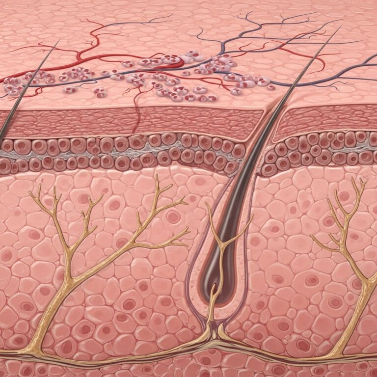
The breast tissue is encircled by a thin but important layer of connective tissue called fascia. This fascia has two distinct layers: the deep layer sits immediately atop the pectoralis muscle, while the superficial layer sits just under the skin. These fascial layers provide structural integrity and separate the breast tissue from underlying and overlying structures.
The skin covering the breast is similar to skin elsewhere on the body, featuring sweat glands, hair follicles, and other normal skin structures. During a comprehensive breast examination, clinicians examine not only the breast tissue itself but also the overlying skin, as changes in skin appearance can indicate underlying pathology.
Blood Supply to the Breast
The breast receives its blood supply primarily from the internal mammary artery, which runs underneath the main breast tissue. This arterial supply provides essential nutrients, particularly oxygen, to support the breast tissue’s metabolic needs and maintain tissue health. Understanding the breast’s vascular anatomy is important for surgical planning and recognizing potential complications.
The blood vessels of the breast appear as thin, tortuous, linear opacities in mammographic imaging. Interestingly, these vessels are more visible in breasts containing more adipose tissue, as the fat allows for better visualization of vascular structures on imaging studies.
Venous Drainage
The venous drainage of the breast follows a dual pathway system. The superficial veins form the venous plexus of Haller, which runs deep to the nipple areolar complex and along the anterior surface of the fascia. The deep veins run alongside the arterial supply of their respective regions. Both superficial and deep veins eventually drain into the internal mammary and axillary veins, which return deoxygenated blood to the central circulation. This efficient venous drainage system prevents blood from pooling in the breast tissue and maintains healthy circulation.
Lymphatic Drainage and Cancer Spread
The lymphatic system of the breast is particularly important in understanding how breast cancer can spread. Lymphatic vessels flow in the opposite direction of blood supply and drain into lymph nodes—specialized structures that filter lymph and help fight infection. Knowledge of lymphatic drainage patterns is crucial for cancer staging and treatment planning.
Lymph Node Groups
Lymph from breast tissue drains into three main groups of lymph nodes: the axillary nodes, the parasternal (internal mammary) nodes, and the supraclavicular nodes. Most lymphatic drainage—approximately 75 percent—flows to the axillary lymph nodes, which are located in the underarm area and serve primarily the lateral quadrants of the breast.
Axillary lymph nodes are classified into three levels based on their location relative to the pectoralis minor muscle. Level I axillary lymph nodes contain three groups and are located inferolateral (below and to the side) to the pectoralis minor muscle. Level II axillary nodes contain one group and are located directly posterior (behind) the pectoralis minor. Level III axillary lymph nodes contain one group and are located superomedial (above and toward the middle) to the pectoralis minor.
Lymphatic vessels draining from the medial quadrants of the breast lead to the internal mammary lymph nodes after passing through intercostal spaces and the pectoralis major muscle. These internal mammary nodes are located immediately medial to the breast tissue and are typically not visible on standard mammograms, though they can be assessed through more advanced imaging or surgical exploration. When breast cancer metastasizes, it usually involves the first lymph node in the chain—called the sentinel lymph node—which surgeons may remove and examine to check for cancer spread.
Associated Muscles and Chest Wall Structures
While the breast itself is primarily composed of glandular and adipose tissue, several muscles of the chest wall are intimately associated with breast anatomy and support its structure.
The Pectoralis Major Muscle
The pectoralis major muscle, the large chest muscle underlying the breast, serves as an important anatomical landmark. On mammographic imaging, the pectoralis major muscle appears as a slightly radiopaque, linear structure located posterior (behind) the breast tissue. During mammography, technicians position patients to include the pectoralis major muscle in the image, as the muscle’s presence helps radiologists determine if adequate breast tissue has been imaged.
Additional Chest Wall Muscles
Several other muscles contribute to the anatomical framework surrounding the breast. The sternalis muscle is a bandlike structure that runs longitudinally along the medial border of the sternum. This muscle can bulge anteriorly when a patient is placed in a decubitus position (lying on their side) and relaxes during imaging. The sternalis muscle can most clearly be seen in craniocaudal (head-to-toe) projection mammograms as an irregular opaque body located medially to the breast.
The external oblique muscle runs along the inferolateral (lower outer) edge of the breast, while the rectus abdominis muscle establishes the inferior (lower) border of the breast. The serratus anterior muscle runs along the lateral aspect of the chest wall and breast, providing additional structural support to the region.
Breast Tissue Composition and Mammographic Appearance
Understanding how different breast tissues appear on mammography helps healthcare providers interpret imaging studies. In mammograms, adipose tissue is radiolucent, meaning it appears relatively dark because it allows X-rays to pass through easily. In contrast, the stroma (fibrous tissue) appears radiopaque, meaning it appears lighter because it absorbs more X-rays. This difference in appearance helps radiologists distinguish between different tissue types and identify abnormalities.
Breast density—the proportion of glandular and fibrous tissue relative to adipose tissue—varies significantly among individuals and can change with age, hormonal status, and other factors. Women with denser breast tissue have more glandular and fibrous tissue relative to fat, which can make mammographic interpretation more challenging and may slightly increase breast cancer risk.
Differences Between Male and Female Breast Anatomy
While the basic anatomical structure of the male and female breast is nearly identical, there is one significant physiological difference. The male breast tissue lacks the specialized lobules present in the female breast, since there is no physiologic need for milk production by males. However, males retain the same basic ductal system and can develop breast disease, including breast cancer, although this occurs far less frequently than in females.
Changes During Pregnancy and Lactation
The breast undergoes dramatic changes during pregnancy and lactation in response to hormonal shifts. During pregnancy, the glandular tissue proliferates, the lobes and lobules expand, and adipose tissue may be replaced by glandular tissue. The areola often darkens, and the nipple may become more prominent. These changes prepare the breast for milk production and delivery. After weaning from breastfeeding, the breast tissue typically returns to its pre-pregnancy state, though complete involution may take several months to years.
Clinical Significance of Breast Anatomy
Understanding breast anatomy is essential for multiple reasons. Healthcare providers use this knowledge to conduct thorough clinical breast examinations, interpret mammographic and ultrasound images, perform surgical procedures, and counsel patients about breast health. Patients benefit from understanding their own anatomy because it helps them recognize normal variations, identify changes that warrant medical attention, and communicate effectively with their healthcare providers about breast concerns.
Frequently Asked Questions
Q: How many lobes does each breast contain?
A: Each breast typically contains approximately 15 to 20 lobes arranged in a radial pattern around the nipple. These lobes vary slightly in size and may not be completely symmetrical between the two breasts.
Q: Why is the axillary tail of Spence clinically important?
A: The axillary tail of Spence is a region of breast tissue extending into the underarm area where breast cancer can develop. Because this tissue may not seem to be located within the main breast, tumors developing there might be overlooked during self-examination, making awareness of this anatomical feature important for early detection.
Q: What are Cooper’s ligaments and what do they do?
A: Cooper’s ligaments are suspensory ligaments that provide structural support to the breast tissue, anchoring it to the underlying pectoralis fascia. They help maintain the breast’s position and shape, but stretching of these ligaments over time can contribute to breast sagging.
Q: Where does breast lymph primarily drain?
A: Approximately 75 percent of breast lymph drains to the axillary (underarm) lymph nodes. Smaller amounts drain to the internal mammary (parasternal) lymph nodes and supraclavicular nodes. Understanding this drainage pattern is crucial for cancer staging and treatment planning.
Q: What is the sentinel lymph node?
A: The sentinel lymph node is the first lymph node in the chain that receives lymphatic drainage from the breast. When breast cancer metastasizes, it typically involves this node first, making sentinel lymph node biopsy an important surgical technique for assessing cancer spread.
References
- Overview of the Breast – Breast Pathology — Johns Hopkins Pathology. Accessed December 1, 2025. https://pathology.jhu.edu/breast/overview
- Breast Anatomy – Radiology Education Resources — UCLA Health, Department of Radiology. Accessed December 1, 2025. https://www.uclahealth.org/departments/radiology/education/breast-imaging-teaching-resources/screening-mammogram/breast-anatomy
- Breast Anatomy and Physiology — National Institute of Health (NIH). National Cancer Institute. https://www.cancer.gov
Similar Articles


Read full bio of medha deb