Bullous Pemphigoid: Essential Guide to Diagnosis & Management
Chronic autoimmune blistering skin disease primarily affecting the elderly: causes, symptoms, diagnosis, and management strategies.

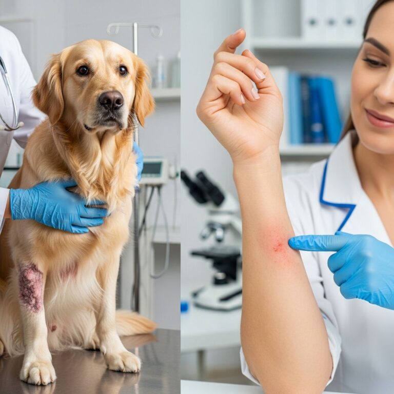
Bullous pemphigoid is a chronic autoimmune skin disorder that predominantly affects older adults, characterized by intensely itchy urticarial plaques, tense blisters, and potential erosions. It arises from autoantibodies targeting hemidesmosomal proteins in the basement membrane, leading to subepidermal separation. This condition, while not life-threatening, significantly impacts quality of life due to severe pruritus and requires long-term management.
What is bullous pemphigoid?
Bullous pemphigoid (BP) represents the most common autoimmune subepidermal blistering disease, with an incidence of 6–30 cases per million per year in those over 60 years. It involves autoantibodies against BP180 (type XVII collagen) and BP230 antigens in hemidesmosomes, disrupting dermo-epidermal adhesion and causing inflammation with eosinophil infiltration.
Unlike pemphigus, blisters in BP are tense and subepidermal, rarely involving mucous membranes. The disease progresses through prodromal (urticarial/eczematous), bullous, and healing phases, often resolving after years but with risk of recurrence.
Who gets bullous pemphigoid?
BP primarily affects individuals over 70 years, with equal prevalence in males and females. Risk factors include neurological conditions (stroke, Parkinson’s, dementia), prior skin trauma, UV therapy, and medications like diuretics, neuroleptics, or PD-1 inhibitors (pembrolizumab, nivolumab) used in cancer therapy. Checkpoint inhibitors induce BP in 0.3% of cases, often after 12 months, featuring prodromal itch and mucosal involvement.
- Peak incidence: 70–80 years
- Risk associations: Neurodegenerative diseases (2–3x higher), psoriasis, lichen sclerosus
- Iatrogenic triggers: Dipeptidyl peptidase-4 inhibitors (gliptins), PD-1/PD-L1 blockers
What causes bullous pemphigoid?
The exact trigger remains unknown, but autoimmunity plays a central role. Genetic factors (HLA-DR4/-DQ7), thermal/physical trauma, and vaccinations have been implicated. In drug-induced cases, culprit agents must be withdrawn promptly. Recent studies highlight epitope spreading, where initial IgG1 antibodies evolve to broader reactivities, exacerbating disease.
Pathophysiology involves complement activation, mast cell degranulation, and eosinophil recruitment, producing proteolytic enzymes that cleave the basement membrane.
What are the clinical features of bullous pemphigoid?
Prodromal phase
Precedes blisters by weeks to months; features severe pruritus with eczematous or urticarial plaques on flexures (axillae, groin), trunk, and extremities. Lesions are symmetric, polymorphic, and may mimic eczema or arthropod bites.
Bullous phase
Tense, dome-shaped bullae (0.5–10 cm) arise on normal or erythematous skin, filled with clear/serous fluid. Blisters resist rupture due to epidermal integrity but may erode if traumatized, leading to crusts and postinflammatory hyperpigmentation. Oral involvement is rare (<20%), unlike mucous membrane pemphigoid.
- Distribution: Flexural limbs, abdomen, groin; spares face/palms/soles
- Pruritus: Intense, sleep-disrupting
- Mucosal: Occasional gingival erosions
Healing phase
Erosions heal without scarring over weeks, leaving hyperpigmentation. Milia may form at blister sites.
Diagnosis of bullous pemphigoid
Clinical suspicion arises from elderly patients with pruritic tense bullae. Confirmation requires biopsy and immunofluorescence.
Skin biopsy
- Perilesional biopsy: Subepidermal blister with eosinophils, neutrophils; festooning of dermal papillae
- Light microscopy: Eosinophilic infiltrate in superficial dermis
Direct immunofluorescence (DIF)
Fishnet linear IgG/C3 deposition along basement membrane zone (BMZ). Gold standard, sensitivity >90%.
Indirect immunofluorescence (IIF)Serum anti-BMZ IgG (titer correlates with severity). Salt-split skin shows roof staining (epidermal side).ELISA
Detects anti-BP180/230 antibodies; >97% specificity for active disease.
| Test | Purpose | Sensitivity/Specificity |
|---|---|---|
| Skin biopsy (H&E) | Histology | 80–90%/High |
| DIF | BMZ deposits | >90%/95% |
| IIF | Serum antibodies | 70–80%/90% |
| BP180 ELISA | Antigen-specific | 80–90%/97% |
Differential diagnosis
- Urticaria: Transient wheals, no blisters
- Pemphigus vulgaris: Flaccid intraepidermal bullae, +Nikolsky sign
- Dermatitis herpetiformis: Grouped vesicles on extensors, IgA DIF
- Linear IgA disease: Linear IgA DIF, annular lesions
- Arthropod bites: History-dependent, resolves spontaneously
- Drug eruptions: Temporal link to medication
BP mimics eczema initially; biopsy differentiates.
Drug-induced bullous pemphigoid
10–15% of cases; common culprits: PD-1 inhibitors (melanoma therapy), gliptins (diabetes), furosemide. Features non-inflammatory phenotype, resolves post-withdrawal but recurs in 30%. Non-blistering forms predominate.
Bullous pemphigoid in children
Rare (<5% cases); mechanobullous variant with trauma-induced blisters on extremities. Benign course, anti-BP180+, resolves spontaneously.
Management and treatment of bullous pemphigoid
Aim: Suppress autoimmunity, halt blisters, relieve itch. Severity-based: localized (<10% BSA) vs. extensive.
Topical therapy (mild disease)
- Clobetasol propionate 0.05% ointment (20–40g/day); controls 70–80% localized cases
- Combine with doxycycline 200mg + nicotinamide 1500–3000mg/day (steroid-sparing)
Systemic corticosteroids (moderate-severe)
Prednisone/prednisolone 0.5–1 mg/kg/day; taper once <3 new blisters/week. Maintenance <10mg/day with adjuvants.
Adjuvant immunosuppressants
- Mycophenolate mofetil (2g/day)
- Azathioprine (2–3mg/kg/day; TPMT screen)
- Methotrexate (7.5–15mg/week)
- Rituximab (refractory; 1g x2, 2 weeks apart)
Dupilumab (anti-IL4/13) emerging for steroid reduction.
Monitoring
Bullous Pemphigoid Disease Area Index (BPDAI) scores activity/pruritus. Track BP, weight, DEXA, glucose, lipids quarterly.
| Severity | First-line | Adjunct/Alternative |
|---|---|---|
| Localized (<10% BSA) | Topical clobetasol | Doxycycline + nicotinamide |
| Moderate (10–20%) | Prednisone 0.5mg/kg + topical | MMF/azathioprine |
| Severe (>20%) | Prednisone 1mg/kg | Rituximab/IVIG |
Complications of bullous pemphigoid
- Infections: Sepsis from eroded skin (mortality 10–40% severe cases)
- Steroid side effects: Osteoporosis, diabetes, hypertension
- Quality of life: Chronic itch, sleep loss, depression
Prevention of bullous pemphigoid
No known prevention; avoid triggers in at-risk patients (e.g., discontinue high-risk drugs).
Outlook and prognosis
Remission in 50–60% after 3–6 years; 20% require lifelong therapy. Mortality 15–25% (age/comorbidities), higher without treatment. Early intervention improves outcomes.
Frequently Asked Questions
What triggers bullous pemphigoid?
Autoimmune response against skin basement membrane; triggered by drugs, trauma, or neurological disease in elderly.
Is bullous pemphigoid contagious?
No, it is autoimmune, not infectious.
How long does treatment last?
Several years; many remit but monitor for relapse.
Does it scar?
Rarely; heals with hyperpigmentation.
Can children get it?
Yes, rarely; milder mechanobullous form.
References
- Bullous pemphigoid – Symptoms, diagnosis and treatment — BMJ Best Practice. 2023. https://bestpractice.bmj.com/topics/en-us/453
- Bullous Pemphigoid – StatPearls — NCBI Bookshelf / D Baigrie. 2023-10-01. https://www.ncbi.nlm.nih.gov/books/NBK535374/
- Bullous pemphigoid — NHS UK. 2023. https://www.nhs.uk/conditions/bullous-pemphigoid/
- Bullous pemphigoid — DermNet NZ. 2024. https://dermnetnz.org/topics/bullous-pemphigoid
- Bullous pemphigoid – Diagnosis and treatment — Mayo Clinic. 2024. https://www.mayoclinic.org/diseases-conditions/bullous-pemphigoid/diagnosis-treatment/drc-20350419
- Bullous Pemphigoid — MSD Manuals Professional Edition. 2024. https://www.msdmanuals.com/professional/dermatologic-disorders/bullous-diseases/bullous-pemphigoid
Similar Articles


Read full bio of Sneha Tete