Burr Holes: Purpose, Procedure, and Recovery
Understanding burr holes: A minimally invasive neurosurgical procedure for brain treatment and monitoring.

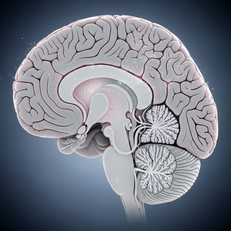
Understanding Burr Holes in Neurosurgery
Burr holes represent a fundamental neurosurgical technique that involves creating small, precise openings in the skull to access the brain and surrounding structures. Unlike more extensive craniotomy procedures that remove larger sections of bone, burr holes are specifically designed for targeted interventions requiring minimal bone disruption. This procedure has become increasingly important in modern neurosurgery, offering neurosurgeons the ability to address various neurological conditions while preserving skull integrity and reducing patient trauma.
The burr hole technique bridges the gap between non-invasive imaging and comprehensive open surgery, providing surgeons with direct access to critical brain structures through carefully positioned small openings. These precise apertures allow for treatment of conditions ranging from acute bleeding around the brain to chronic neurological disorders requiring fluid management.
What Are Burr Holes?
Burr holes are small openings created in the skull bone using specialized surgical instruments designed for this purpose. The term “burr” refers to the rotating cutting tool surgeons use to carefully perforate the bone. Typically ranging from 14 to 20 millimeters in diameter, these openings are much smaller than traditional craniotomy incisions, which can measure several centimeters across.
The procedure involves several layers of precision work. Surgeons first make an incision in the scalp, then use specialized bone-cutting instruments to carefully create the opening through the skull’s outer and inner tables. The size and location of each burr hole are meticulously planned based on the patient’s specific condition and the surgical objectives.
What distinguishes burr holes from other neurosurgical approaches is their minimally invasive nature combined with their effectiveness. The procedure allows surgeons to access brain structures, remove accumulated fluid, place monitoring devices, or insert therapeutic implants without requiring extensive bone removal or the extended recovery period associated with larger surgical approaches.
Common Conditions Treated with Burr Holes
Subdural Hematoma
One of the most frequent indications for burr hole placement is the treatment of subdural hematomas, which represent bleeding in the space between the brain and the outermost membrane surrounding it. These hematomas typically result from head trauma, falls, or sometimes minor head injuries, particularly in older adults or those taking blood-thinning medications. The accumulation of blood creates pressure on the brain tissue, potentially causing neurological deterioration if not promptly addressed. Burr holes allow neurosurgeons to access and evacuate this blood collection effectively.
Cerebrospinal Fluid Disorders
Burr holes play a crucial role in managing various cerebrospinal fluid (CSF) disorders, including hydrocephalus and other conditions affecting fluid circulation around the brain. Surgeons use burr holes to insert ventriculoperitoneal shunts, which divert excess CSF away from the brain and into the abdominal cavity where it can be reabsorbed. Additionally, burr holes provide access for endoscopic third ventriculostomy procedures, which create alternative pathways for CSF circulation when normal drainage pathways are blocked.
Epidural Hematoma
Similar to subdural hematomas, epidural hematomas involve bleeding in a different anatomical space—between the skull and the dura mater membrane. Burr holes provide effective access for evacuating this blood collection and controlling the source of bleeding, particularly when arterial vessels are involved in the hemorrhage.
Brain Biopsy
In cases where tissue diagnosis is required, surgeons may use burr holes to obtain brain tissue samples for pathological examination. This approach helps diagnose tumors, infections, or other infiltrative brain diseases while minimizing damage to surrounding brain tissue.
Intracranial Pressure Monitoring
Burr holes serve as access points for inserting intracranial pressure (ICP) monitoring devices in critically ill patients. These devices measure pressure inside the skull, helping clinicians guide treatment decisions for patients with severe brain injuries, infections, or other conditions affecting intracranial dynamics.
The Burr Hole Procedure
Preoperative Preparation
Before undergoing a burr hole procedure, patients undergo comprehensive preoperative evaluation including detailed imaging studies such as computed tomography (CT) scans or magnetic resonance imaging (MRI). These imaging studies help neurosurgeons precisely locate the target pathology and determine optimal burr hole placement. Patients receive standard preoperative testing including blood work and cardiac evaluation as deemed necessary.
Patients are typically instructed to fast for several hours before surgery and receive information about anesthesia and the general operative plan. Clear communication between the surgical team and patient helps establish realistic expectations about the procedure.
Operative Technique
The burr hole procedure begins with general anesthesia to ensure patient comfort and safety throughout the operation. The surgical team then prepares and sterilizes the operative field using standard antiseptic techniques. Following scalp incision over the planned burr hole location, the surgeon carefully separates the muscle and fascia layers to expose the skull bone.
Using specialized surgical instruments including perforator drills with specific burr bits, the surgeon creates the opening through the skull bone. Standard 14-millimeter burr holes and expanded 20-millimeter burr holes represent common sizes, with the choice depending on the specific procedure being performed. The surgeon must carefully control the depth of drilling to penetrate both the outer and inner tables of the skull while avoiding inadvertent injury to the dura mater and underlying brain tissue.
Once the burr hole is created, the dura mater may be opened depending on the specific procedure. Surgeons then perform the intended therapeutic intervention, whether evacuating hematoma, placing a shunt catheter, obtaining biopsy tissue, or inserting a monitoring device. Following completion of the therapeutic objective, layers are closed in reverse anatomical order, with careful attention to hemostasis and watertight closure when appropriate.
Advanced Imaging Applications
Recent innovations in burr hole technology include the placement of sonolucent burr hole covers—specially designed implants that allow transcranial ultrasound imaging through the burr hole site. These covers enable radiation-free postoperative imaging of ventricular size and brain structures, offering an alternative to repeated CT or MRI studies. This development represents a significant advancement in postoperative monitoring capabilities for patients with cerebrospinal fluid disorders, particularly in reducing cumulative radiation exposure.
Recovery and Postoperative Course
Immediate Postoperative Period
Following burr hole surgery, patients typically spend time in the recovery room for close monitoring before transfer to an inpatient unit. The initial postoperative period involves regular assessments of neurological function, vital signs, and wound condition. Pain management is addressed with appropriate medications, and patients receive instructions about activity restrictions and wound care.
Expected Recovery Timeline
Recovery following burr hole procedures is generally faster than recovery after more extensive neurosurgery. Most patients experience mild to moderate discomfort at the incision site, which typically resolves within days to weeks. Patients may resume normal activities gradually as tolerated, though specific activity restrictions depend on the underlying condition and the specific procedure performed.
Bone Regrowth Considerations
An interesting aspect of burr hole healing involves natural bone regrowth at the burr hole site. Research demonstrates that bone regrowth after burr hole craniostomy is common, occurring rapidly within the first 6 to 12 months and subsequently stabilizing. On average, the burr hole closes approximately 25 percent at 6 months, with minimal additional closure over the following 18 months. The mean residual diameter for standard 14-millimeter burr holes stabilizes around 9.4 millimeters, while expanded 20-millimeter burr holes stabilize around 15.4 millimeters. Notably, bone regrowth rates are not associated with patient characteristics including age, sex, skull thickness, or the underlying cause of cerebrospinal fluid disorder.
This natural bone healing process reflects the body’s remarkable capacity to restore structural integrity while maintaining the functional benefits achieved through the surgical intervention. The gradual closure does not typically compromise the effectiveness of therapeutic devices like shunts or interfere with the primary surgical objective.
Risks and Complications
Infection
Like any surgical procedure, burr holes carry a risk of infection at the incision site or potentially of meningitis affecting the brain membranes. Strict sterile technique and appropriate prophylactic antibiotics minimize this risk substantially. Patients should report signs of infection such as fever, increasing redness or drainage at the incision site, or severe headache.
Hemorrhage
Bleeding complications can occur during or after the procedure. While the burr hole technique minimizes vascular injury risk compared to larger procedures, bleeding can still happen. Surgeons take precautions to identify and control bleeding sources before closure. Postoperative monitoring helps detect delayed bleeding requiring intervention.
Neurological Injury
Although burr holes minimize brain trauma compared to larger procedures, injury to brain tissue, blood vessels, or nerves is possible. Careful preoperative planning and surgical technique significantly reduce this risk. Any new or worsening neurological symptoms after surgery should be promptly reported to the surgical team.
Cerebrospinal Fluid Leak
If the dura mater is opened during the procedure, CSF leakage can occasionally occur. Careful closure techniques help prevent this complication. Small CSF leaks typically resolve spontaneously, while larger leaks may require intervention.
Device-Related Complications
When burr holes are used for shunt placement or other device insertion, complications specific to the implanted device can occur, including obstruction, malposition, or malfunction. Regular follow-up imaging helps identify such issues early.
Burr Holes Versus Other Neurosurgical Approaches
| Feature | Burr Holes | Craniotomy | Endoscopy |
|---|---|---|---|
| Incision Size | Very small (1-2 cm) | Large (5-10 cm) | Minimal |
| Bone Removal | Small openings | Large bone flap | None |
| Access Capability | Targeted access | Extensive access | Limited access |
| Recovery Time | Faster | Slower | Very fast |
| Tissue Trauma | Minimal | Moderate to significant | Very minimal |
Frequently Asked Questions
How long does a burr hole procedure typically take?
The duration varies depending on the specific procedure being performed. Simple burr hole placement for hematoma evacuation might take 30 to 60 minutes, while more complex procedures such as endoscopic third ventriculostomy or multiple burr hole placements may require longer operative times. The surgical team can provide specific time estimates based on the planned procedure.
Will I have a permanent scar from burr hole surgery?
A small scar typically remains at the incision site, though it usually becomes less noticeable over time. The size is generally much smaller than scars from extensive neurosurgery. Hair typically covers the scar well in most patients, and over time, the scar becomes increasingly inconspicuous.
Can burr holes be used for emergency treatment?
Yes, burr holes are frequently used for emergency treatment of acute conditions such as epidural or subdural hematomas causing neurological deterioration. The technique’s relative simplicity and speed make it well-suited for emergency situations requiring rapid access to the intracranial space.
What happens if the burr hole fills in completely with new bone?
While bone regrowth does occur, complete closure is uncommon, and even partial closure does not typically compromise the effectiveness of shunts or other therapeutic interventions. Some patients undergo follow-up procedures, but most do not require intervention related to bone regrowth.
Are burr holes reversible or permanent?
While the initial burr hole is permanent, bone regrowth gradually fills the opening. If future surgery through the same location becomes necessary, surgeons can typically create a new burr hole or access through the existing site, as the bone regrowth does not completely eliminate the pathway.
How is pain managed after burr hole surgery?
Pain management typically involves prescription medications in the immediate postoperative period, with transition to over-the-counter pain relievers as healing progresses. Most patients experience mild to moderate discomfort that resolves within several weeks. The surgical team provides specific pain management instructions and monitors the patient’s comfort level.
References
- Craniotomy: What is it, and is it different from craniectomy? — Medical News Today. https://www.medicalnewstoday.com/articles/craniotomy
- Bone Regrowth After Frontal Burr Hole Craniostomy — National Institutes of Health, NIH. 2024. https://pmc.ncbi.nlm.nih.gov/articles/PMC11810037/
- Bone Regrowth After Frontal Burr Hole Craniostomy: Natural History — Johns Hopkins University, Neurosurgery Practice. September 10, 2024. https://pure.johnshopkins.edu/en/publications/bone-regrowth-after-frontal-burr-hole-craniostomy-natural-history/
- First Experience With Postoperative Transcranial Ultrasound Through Sonolucent Burr Hole Covers — Neurosurgery, Johns Hopkins University School of Medicine. 2023. https://pubmed.ncbi.nlm.nih.gov/36637272/
Similar Articles


Read full bio of medha deb