Buruli Ulcer: Complete Guide To Symptoms, Diagnosis & Care
Understanding Buruli ulcer: causes, symptoms, diagnosis, treatment, and prevention of this neglected tropical skin disease caused by Mycobacterium ulcerans.

What is Buruli ulcer?
Buruli ulcer, also known as Bairnsdale ulcer or Mycobacterium ulcerans infection, is a chronic, progressive skin disease classified as a neglected tropical disease by the World Health Organization (WHO). It is caused by the bacterium Mycobacterium ulcerans, which produces a potent toxin called mycolactone. This toxin destroys skin cells, small blood vessels, and subcutaneous fat, leading to painless ulcers and extensive tissue necrosis.
The disease primarily affects the skin but can involve deeper tissues, including bone, causing deformities and permanent disability if not treated early. Buruli ulcer is most common in tropical and subtropical regions, with over 3,000 cases reported annually worldwide, though underreporting is significant due to its occurrence in remote areas.
Without intervention, lesions enlarge over weeks to months, resulting in large ulcers that heal with scarring. Early diagnosis is crucial to minimize skin loss and prevent complications like secondary infections, osteomyelitis, or functional limitations.
Who gets Buruli ulcer?
Buruli ulcer predominantly affects children under 15 years, comprising about 50% of cases, but can occur at any age. It is more common in rural, marshy, or wetland areas where environmental conditions favor bacterial growth. High-risk populations include those engaged in farming, fishing, or gardening near stagnant water bodies.
Outbreaks have been reported in West Africa (e.g., Ghana, Benin), Australia (Victoria), and parts of Japan, but sporadic cases occur globally. Travelers to endemic areas are at low risk but should remain vigilant.
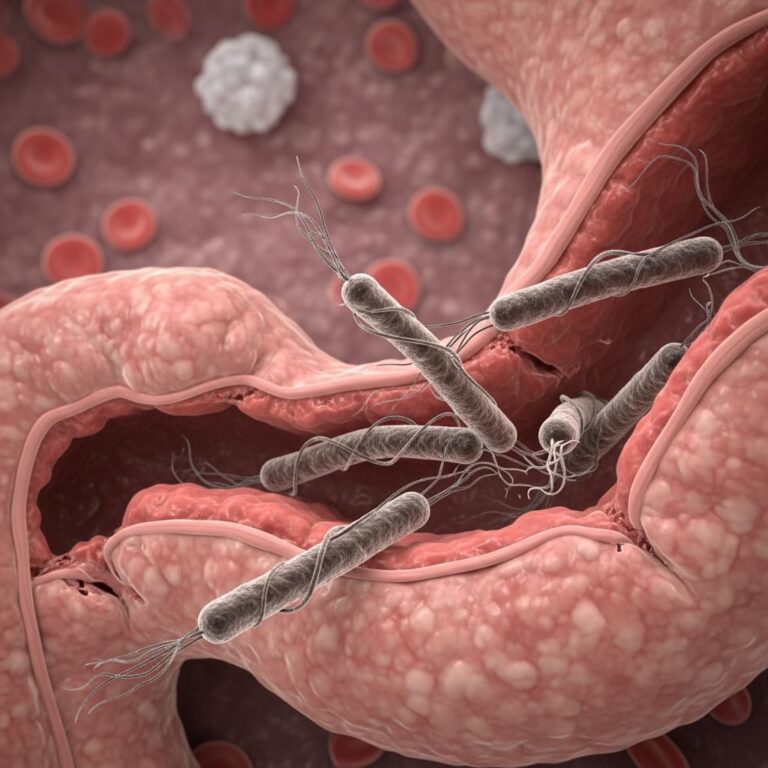
Causes
The primary causative agent is Mycobacterium ulcerans, a slow-growing pathogen related to Mycobacterium tuberculosis and M. leprae. The bacterium thrives in aquatic environments and produces mycolactone, an immunosuppressant toxin responsible for tissue destruction without invoking a strong inflammatory response, explaining the painless nature of lesions.
Multiple strains exist, influencing transmission patterns and virulence. The infection leads to local cell death (necrosis) rather than systemic illness, distinguishing it from other mycobacterial diseases.
Transmission
The exact mode of transmission remains unknown, despite extensive research. There is no evidence of direct human-to-human spread. Hypotheses include:
- Traumatic inoculation via skin breaks contaminated with water, mud, or vegetation from endemic areas.
- Vector transmission by aquatic insects like Naucoridae water bugs or mosquitoes, supported by observational data from outbreaks.
- Consumption of contaminated water, though unproven.
Incubation periods range from 1 to 9 months (average 4-5 months), longer than most bacterial infections, complicating epidemiological tracing.
Clinical features of Buruli ulcer
Buruli ulcer evolves through stages, often starting asymptomatically. Early lesions mimic insect bites, progressing slowly without pain or fever.
Pre-ulcerative stage
Initial presentations include:
- Nodule: Firm, painless subcutaneous lump 1-2 cm in diameter, common on limbs.
- Plaque: Indurated, flat elevated area with irregular borders.
- Oedema: Diffuse swelling, often on legs or arms, rarely face.
These may persist for weeks to months before ulcerating.
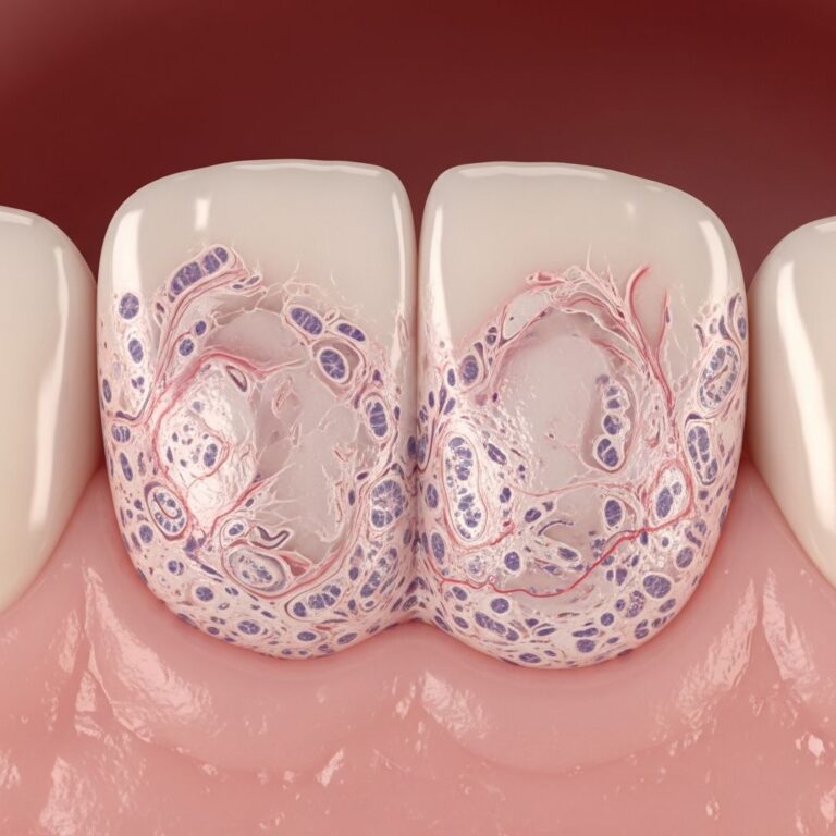
Ulcerative stage
Ulcers form centrally with undermined edges, featuring characteristic white, necrotic slough. They enlarge progressively (up to 15+ cm), remaining painless unless secondarily infected. Sites: 35% lower limbs, 30% upper limbs, 20% trunk.
WHO Categories:
| Category | Description | Prevalence |
|---|---|---|
| I | Single small lesion <5 cm diameter | 32% |
| II | Plaques, oedematous or ulcerative 5-15 cm | 35% |
| III | >15 cm, disseminated, osteomyelitis | 33% |
Complications
Untreated, ulcers cause:
- Extensive tissue loss and contractures.
- Bone/joint involvement (osteomyelitis).
- Secondary bacterial infections, fever, swelling.
- Chronic disability: 25-50% of cases without treatment.
Diagnosis
Diagnosis combines clinical suspicion and laboratory confirmation, especially in non-endemic areas.
- Clinical: History of exposure to endemic areas, painless enlarging ulcer.
- Swabs/Biopsy: For microscopy (acid-fast bacilli), culture (gold standard but slow, 8-12 weeks), histopathology (necrosis, minimal inflammation).
- PCR: Detects M. ulcerans DNA rapidly, highly sensitive.
- IS2404 PCR: Specific insertion sequence for confirmation.
Differential diagnoses: fungal infections, tuberculosis, leishmaniasis, pyoderma. Early biopsy prevents misdiagnosis.
Treatment
WHO-recommended regimen: 8-week combination antibiotics, shifting from streptomycin/rifampicin to rifampicin (10 mg/kg daily) + clarithromycin (7.5 mg/kg twice daily) for efficacy and reduced injectables.
- Small lesions (Category I): Antibiotics alone often heal completely.
- Large lesions: Add surgery (debridement, grafting) + wound care.
- Adjuncts: Painless dressings, physiotherapy, rehabilitation.
Success rates: >90% cure without surgery if early; recurrence rare post-treatment.
Prevention
No vaccine exists. Strategies focus on exposure reduction:
- Wear long clothing, gloves during outdoor work.
- Apply DEET insect repellent, avoid dusk/dawn in wetlands.
- Protect/cover wounds; wash soil/water exposure promptly.
- Community surveillance for early detection.
Frequently Asked Questions
What is the incubation period for Buruli ulcer?
The incubation period ranges from 1 to 9 months, averaging 4-5 months from exposure to lesion appearance.
Is Buruli ulcer contagious?
No, there is no person-to-person transmission.
Can Buruli ulcer be cured?
Yes, with prompt antibiotics and supportive care, cure rates exceed 90%, preventing disability.
How do I know if my skin lesion is Buruli ulcer?
See a doctor for painless, enlarging nodules/ulcers after exposure to endemic areas; confirm with PCR or biopsy.
Where is Buruli ulcer most common?
Africa (Ghana, Benin), Australia (Victoria), and parts of Asia-Pacific.
References
- Buruli ulcer | Better Health Channel — Victoria Government. 2023. https://www.betterhealth.vic.gov.au/health/healthyliving/Buruli-ulcer
- Buruli Ulcer: Causes, Diagnosis, and More — WebMD. 2024-01-15. https://www.webmd.com/a-to-z-guides/what-is-buruli-ulcer
- Buruli ulcer | About Neglected Tropical Diseases — Eisai Co. 2023. https://www.eisai.com/sustainability/atm/ntds/diseases/buruli.html
- Buruli ulcer (Mycobacterium ulcerans infection) — World Health Organization. 2024-05-22. https://www.who.int/news-room/fact-sheets/detail/buruli-ulcer-(mycobacterium-ulcerans-infection)
- Buruli Ulcer: a Review of the Current Knowledge — PMC (NCBI). 2018-11-20. https://pmc.ncbi.nlm.nih.gov/articles/PMC6223704/
- Buruli Ulcer – Control of Communicable Diseases Manual — APHA Publications. 2025. https://ccdm.aphapublications.org/doi/10.2105/CCDM.2745.038
Similar Articles


Read full bio of medha deb