Cartilage Regeneration: Advanced Treatment Options
Explore innovative cartilage regeneration techniques and therapies for joint repair.

Understanding Cartilage Regeneration
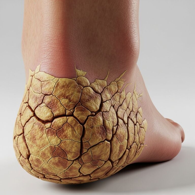
Cartilage damage represents one of the most challenging orthopedic conditions to treat due to the tissue’s inherent limitations in self-healing. Unlike other tissues in the body, articular cartilage possesses minimal regenerative capacity, meaning injuries often lead to progressive deterioration and eventual osteoarthritis if left untreated. The surface layer of cartilage lacks blood vessels and nerves, which severely restricts the body’s natural healing mechanisms. This fundamental problem has driven researchers and clinicians to develop innovative cartilage regeneration strategies that leverage advanced biomaterials and cellular therapies.
The field of cartilage regeneration has evolved dramatically over the past two decades, moving from simple palliative treatments to sophisticated biological solutions. At Johns Hopkins Medicine, the Translational Tissue Engineering Center (TTEC) and the Center for Musculoskeletal Research have pioneered groundbreaking approaches that combine tissue engineering principles with clinical applications. These advances offer hope to millions of patients suffering from cartilage defects, whether caused by trauma, degenerative disease, or congenital conditions.
The Challenge of Articular Cartilage Repair
Articular cartilage, the smooth tissue that covers the ends of bones in joints, faces unique challenges when damaged. This specialized tissue is composed of chondrocytes (cartilage cells) embedded in an extracellular matrix rich in collagen and proteoglycans. When injury occurs, the cartilage’s avascular nature means it cannot recruit immune cells or growth factors necessary for healing. Additionally, chondrocytes have limited mitotic activity, so they cannot rapidly divide to replace damaged tissue.
Traditional approaches to cartilage repair, including osteochondral transplants and cartilage grafts, have demonstrated limited long-term success. These methods often fail due to poor integration with surrounding tissue, inadequate vascularization, and progressive degeneration of transplanted material. The quest for more effective solutions has led researchers to explore biomaterials and regenerative medicine strategies that address the fundamental biological requirements for cartilage healing.
Hydrogel Scaffolding Technology
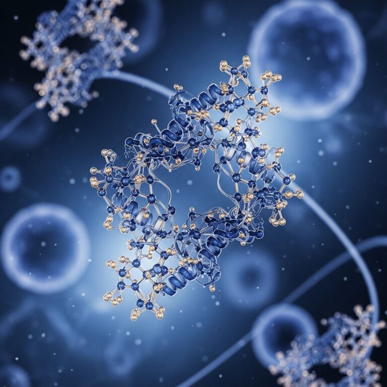
One of the most promising advances in cartilage regeneration is the development of hydrogel scaffolding systems. Researchers at Johns Hopkins University developed a poly(ethylene glycol) diacrylate (PEGDA) hydrogel specifically designed to support cartilage matrix production while maintaining ease of surgical application. This innovative biomaterial serves as a three-dimensional scaffold that mimics the natural extracellular matrix, providing structural support for newly forming cartilage tissue.
Clinical Evidence for Hydrogel Success
A landmark clinical study published in Science Translational Medicine demonstrated the effectiveness of hydrogel scaffolding combined with standard microfracture surgery. In this pilot study, 15 patients with focal cartilage defects of the knee received hydrogel implants during microfracture surgery, while three control patients underwent microfracture alone. The results were compelling: patients receiving hydrogel implants achieved significantly higher levels of tissue fill, with magnetic resonance imaging showing new cartilage filling an average of 86 percent of the defect compared to 64 percent in control patients.
Beyond tissue growth, patients with hydrogel implants reported substantially greater pain reduction in the six months following surgery. Magnetic resonance spin-spin relaxation times (T2) measurements demonstrated decreasing water content and increased tissue organization over time in treated patients, indicating maturation and improved quality of regenerated cartilage. Importantly, no major adverse events were observed during the study period, establishing the safety profile of this biomaterial approach.
How Hydrogel Scaffolding Works
The hydrogel system functions through multiple complementary mechanisms. The squishy hydrogel material is implanted during microfracture surgery, in which tiny holes are punched in the bone near the injured cartilage. These microfractures stimulate the patient’s own specialized stem cells to emerge from bone marrow and migrate to the injury site. The hydrogel provides a nurturing microenvironment that supports and nourishes the healing process, allowing these stem cells to differentiate into chondrocytes and produce cartilage matrix.
To ensure the hydrogel remains in place and properly integrated with the surrounding tissue, researchers developed a chondroitin sulfate adhesive that covalently bonds the hydrogel to cartilage and bone tissue in articular defects. This combination of hydrogel and adhesive represents a practical biomaterials strategy with demonstrated clinical potential for improving articular cartilage defect treatment.
Stem Cell-Based Regeneration Approaches
Complementary to hydrogel technology, stem cell therapies represent another frontier in cartilage regeneration. Muscle-derived stem cells (MDSCs) have emerged as particularly promising candidates for cartilage repair applications. These cells can differentiate into multiple cell types, including chondrocytes, and can be genetically engineered to produce trophic factors essential for cartilage growth and stability.
Genetic Engineering for Enhanced Regeneration
Research has demonstrated that MDSCs genetically engineered to express bone morphogenic protein-4 (BMP-4) show dramatically enhanced cartilage regeneration capacity. In preclinical studies, transfected MDSCs produced significantly more Type II collagen-producing colonies and demonstrated dense extracellular matrix formation compared to non-transfected cells. When implanted into full-thickness articular cartilage defects in animal models, transfected MDSCs showed chondrocyte differentiation with robust and durable cartilage matrix production, with histologic appearance indistinguishable from normal hyaline cartilage up to 24 weeks after implantation.
In contrast, non-transfected MDSCs demonstrated little to no chondrocyte differentiation, progressive thinning, and a lack of bonding to adjacent undamaged cartilage. This dramatic difference underscores the importance of engineering cellular therapies to maximize regenerative potential.
Microfracture Surgery: The Foundation
Microfracture surgery forms the foundational technique upon which many cartilage regeneration strategies are built. This minimally invasive procedure involves creating small holes or fractures in the subchondral bone beneath the cartilage defect. The controlled trauma stimulates the migration of bone marrow-derived mesenchymal stem cells to the injury site, where they can differentiate into cartilage-producing cells.
While microfracture alone provides some benefit, the addition of biological scaffolds and growth factors significantly enhances the quantity and quality of regenerated tissue. This synergistic approach combines mechanical stimulation with biological support, creating optimal conditions for cartilage repair.
Biomaterial Innovation and Future Directions
The success of initial hydrogel and adhesive systems has inspired continued innovation in biomaterial design. Researchers at Johns Hopkins are developing next-generation implants in which the hydrogel and adhesive will be combined into a single, unified material. This advancement would simplify surgical application and potentially improve integration and efficacy.
Beyond cartilage regeneration itself, scientists are exploring complementary technologies to address the broader challenges of joint disease. Current research efforts include technologies to lubricate joints and reduce inflammation, recognizing that successful cartilage repair must occur within a healthy joint environment.
Research Infrastructure and Collaboration
The success of cartilage regeneration research at Johns Hopkins reflects the institution’s commitment to collaborative, interdisciplinary investigation. The Center for Musculoskeletal Research brings together basic, translational, and clinical scientists focused on understanding musculoskeletal conditions. This diverse team, composed of experienced faculty from multiple departments, creates an environment where fundamental discoveries can be rapidly translated into clinical applications.
The Translational Tissue Engineering Center (TTEC), directed by Professor Jennifer Elisseeff, exemplifies this translational approach. Dr. Elisseeff and her colleagues have systematically advanced hydrogel technology from laboratory testing to animal studies and ultimately to human clinical trials. This methodical progression ensures that new therapies are thoroughly validated before clinical application.
Clinical Applications and Patient Benefits
Cartilage regeneration therapies address a significant clinical need. Patients with cartilage defects often experience chronic pain, limited mobility, and reduced quality of life. The development of effective regeneration strategies offers the potential to restore joint function, reduce pain, and slow or prevent progression to osteoarthritis.
Current applications focus primarily on focal cartilage defects of the knee, particularly on the medial femoral condyle, where microfracture is technically feasible and outcomes can be readily assessed. However, the principles underlying these regeneration strategies may be adapted for other joints and broader applications in joint disease management.
Regulatory Pathway and Future Clinical Translation
The clinical validation of hydrogel technology continues to advance through ongoing clinical trials managed in collaboration with commercial partners. These expanded trials aim to generate the safety and efficacy data necessary for regulatory approval in the United States and Europe. The systematic approach to clinical translation reflects the rigorous standards required for new medical devices and biologics in modern healthcare.
Frequently Asked Questions
Q: Why is cartilage so difficult to repair naturally?
A: Articular cartilage lacks blood vessels, nerves, and abundant cells necessary for self-healing. This avascular nature prevents the body from recruiting immune cells and growth factors needed for tissue repair, making spontaneous healing extremely limited.
Q: How does hydrogel scaffolding improve cartilage repair?
A: The hydrogel provides a three-dimensional structure that mimics the natural cartilage matrix, supporting and nourishing stem cells that migrate from bone marrow following microfracture. This biological environment promotes chondrocyte differentiation and cartilage matrix production.
Q: What are muscle-derived stem cells and why are they useful?
A: Muscle-derived stem cells are multipotent cells that can differentiate into multiple cell types, including chondrocytes. When genetically engineered to express growth factors like BMP-4, they demonstrate enhanced ability to regenerate cartilage tissue.
Q: Is microfracture surgery still used if I receive hydrogel treatment?
A: Yes, microfracture remains the foundation of current regeneration strategies. The hydrogel scaffold is implanted during the microfracture procedure to enhance the body’s natural healing response stimulated by the controlled bone fractures.
Q: Are there any side effects or complications from these treatments?
A: Clinical studies have shown no major adverse events associated with hydrogel implants over the study periods evaluated. However, as with any surgical procedure, patients should discuss potential risks with their orthopedic surgeon.
Q: When will these advanced therapies be widely available?
A: Clinical trials continue to expand the evidence base for regulatory approval. The timeline for widespread clinical availability depends on successful completion of ongoing studies and regulatory clearance processes.
References
- TTEC clinical study proves successful in cartilage repair — Johns Hopkins University Biomedical Engineering. 2013-01-09. https://www.bme.jhu.edu/news-events/news/ttec-clinical-study-proves-successful-in-cartilage-repair/
- Tissue engineers use new biomaterial to fix knee cartilage — Johns Hopkins University Hub/Gazette. 2013-02. https://hub.jhu.edu/gazette/2013/february/tissue-engineers-biomaterial-knee-repair/
- Human cartilage repair with a photoreactive adhesive-hydrogel — PubMed/National Institutes of Health. 2013. https://pubmed.ncbi.nlm.nih.gov/23303605/
- Effect of Muscle-Derived Stem Cells on Cartilage Repair — Johns Hopkins Arthritis Center. https://www.hopkinsarthritis.org/arthritis-news/effect-of-muscle-derived-stem-cells-on-cartilage-repair/
- Center for Musculoskeletal Research at Johns Hopkins University — Johns Hopkins University. https://hopkinscmr.jhmi.edu
Similar Articles


Read full bio of medha deb