Cavernous Malformation: Symptoms, Causes & Treatment
Understanding cavernous hemangiomas: causes, symptoms, diagnosis and treatment options for brain lesions.
What Is a Cavernous Malformation?
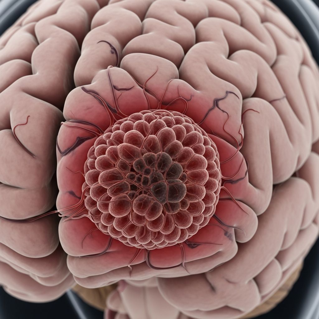
A cavernous malformation, commonly referred to as a cavernoma or cavernous hemangioma, is an abnormal tangle of thin-walled blood vessels. These lesions typically develop in the brain, brainstem, and spinal cord, though they can occasionally occur in other parts of the body. The distinctive appearance of these malformations resembles a raspberry, with blood-filled spaces separated by connective tissues. What makes these lesions particularly concerning is their structural weakness—the thin walls of these abnormal blood vessels lack adequate support compared to normal capillaries, making them significantly more prone to leaking blood.
The size of cavernous hemangiomas varies considerably, ranging from a fraction of an inch to dime-sized lesions or larger. Despite their fragile nature, many people with these malformations experience few to no problems throughout their lives. However, when bleeding occurs, it can lead to serious complications including seizures, paralysis, or stroke. The severity of these complications depends on multiple factors unique to each individual case.
Common Names and Terminology
Healthcare providers use several different terms to describe cavernous malformations, which can sometimes create confusion for patients. Understanding these alternative names helps you communicate more effectively with your medical team:
- Cavernous hemangioma
- Cavernoma
- Cerebral cavernous malformation
- CCM (abbreviation for cerebral cavernous malformation)
- Cavernous angioma
- Familial cavernous malformation
Risk Factors and Who Is Most Affected
Certain individuals face a higher risk of developing cerebral cavernous malformations. Your risk may be elevated if you have a family history of cavernomas, as genetic factors play a significant role in their development. People with a hereditary predisposition are more likely to develop multiple malformations throughout their brain and spinal cord. Additionally, certain genetic variants can directly cause the formation of these lesions, which healthcare providers can identify through genetic testing.
Age is another consideration—symptoms typically appear between ages 20 and 60, though cavernomas can develop at any age. Some individuals may have had these lesions since childhood without experiencing noticeable symptoms until adulthood. Understanding your personal risk profile helps guide preventive care and monitoring strategies.
Symptoms of Cavernous Malformations
Many people with cavernous malformations experience no symptoms at all and may never realize they have the condition. However, when symptoms do occur, they typically indicate that bleeding has happened within the malformation. Common symptoms include:
- Headaches
- Seizures
- Dizziness or vertigo
- Vision problems or blurred vision
- Weakness or numbness in limbs
- Balance difficulties
- Difficulty with coordination
- Speech problems or slurred speech
- Sensory disturbances
The severity and type of symptoms vary based on the size and location of the malformation. A lesion in a critical brain area may cause more pronounced symptoms than one in a less sensitive region. Importantly, you might not experience any symptoms during an actual bleed, which is why regular monitoring through imaging is essential for those with known cavernomas.
Understanding the Risk of Bleeding
One of the most important statistics for patients to understand is the annual bleeding risk. Research estimates that if you have a cavernous malformation, you have approximately a 2.4% chance of experiencing a bleed in any given year. While this may seem like a relatively low percentage, the cumulative risk increases over time, particularly for individuals with multiple malformations or those with a history of previous bleeding.
Related Stories



Several factors determine whether your particular cavernous malformation poses significant danger, including its location in the brain, its size, whether it has previously bled, and your overall neurological health. Each person’s situation is unique, which is why personalized risk assessment by your healthcare provider is crucial for developing an appropriate management plan.
Diagnosis of Cavernous Malformations
Healthcare providers diagnose cerebral cavernous malformations through a combination of physical examination and advanced imaging technology. During your initial consultation, your provider will review your medical history and any symptoms you’ve been experiencing. They will perform a thorough neurological examination to assess your cognitive function, balance, and other relevant neurological parameters.
Imaging Studies
Magnetic Resonance Imaging (MRI) is the gold standard for visualizing cavernous malformations and identifying any evidence of bleeding or swelling. For more detailed assessment, your provider may order a special type of MRI scan called susceptibility-weighted imaging (SWI), which is considerably more sensitive than conventional MRI. This advanced imaging technique can detect even the tiniest cavernous hemangiomas and can reveal remnants of previous bleeds that might not be visible on standard scans. This information helps your care team assess your individual risk for future complications.
Genetic Testing
Your provider may recommend genetic testing to identify whether a specific genetic variant caused your cavernoma. This is particularly important if you have a family history of cavernous malformations or if you have multiple lesions. Identifying the genetic cause can help predict the likelihood of developing additional malformations and can inform family members about their own risk.
When Are Cavernous Malformations Diagnosed?
Typically, healthcare providers do not diagnose cerebral cavernous malformations until they rupture and cause bleeding that produces noticeable symptoms. However, some people with asymptomatic cavernomas discover the condition incidentally when undergoing brain imaging for an unrelated reason, such as evaluating a different neurological concern or following a head injury.
Treatment Options for Cavernous Malformations
Treatment approaches for cavernous malformations vary significantly based on the specific characteristics of your lesion, including its location within the brain or spinal cord, whether it is currently bleeding, and whether it is causing symptoms.
Observation and Monitoring
If your cavernoma is not bleeding and is not causing symptoms, your healthcare provider may recommend a conservative approach involving regular imaging scans to monitor the lesion over time. During this surveillance period, you will learn to recognize warning signs and understand when to contact your healthcare provider urgently if symptoms develop. This approach allows you to maintain quality of life while ensuring that any changes in your condition are detected promptly.
Medication Management
For individuals experiencing symptoms such as seizures related to their cavernous malformation, medications can help manage these symptoms effectively. Anti-seizure medications are commonly prescribed to reduce the frequency and severity of seizures without requiring surgical intervention. Your provider will work with you to find the medication regimen that provides optimal symptom control while minimizing side effects.
Surgical Treatment
Your healthcare provider may consider surgical removal of the cavernous malformation if certain conditions are met. Surgery may be recommended when:
- The malformation is actively bleeding or has bled previously
- It is causing progressive neurological symptoms
- It is located in an area of the brain that is surgically accessible
- The benefit of removal outweighs the surgical risks
- It is causing intractable seizures that cannot be controlled with medication
The outcome of removing a cerebral cavernous malformation is usually successful when your surgeon can safely access and remove it. However, surgery carries inherent risks, which your care team will discuss thoroughly before proceeding. The decision to pursue surgical intervention requires careful consideration of your individual circumstances and close collaboration with your medical team.
Cavernous Malformations Versus Arteriovenous Malformations
Cavernous malformations and arteriovenous malformations (AVMs) are both vascular lesions that can occur in the brain and spinal cord, and they can appear similar in size depending on the number of blood vessels involved. Both types can rupture and cause bleeding. However, there are important differences between these conditions.
AVMs involve high-pressure blood flow through abnormal vessel connections, which means that when they bleed, the hemorrhage tends to be more severe and causes more extensive damage than bleeding from a cavernous malformation. After AVMs, cavernous malformations are the most likely type of blood vessel lesion to require treatment. While AVMs are typically treated with various specialized approaches, cavernous malformations are generally best treated with surgery to remove the hemangioma when intervention is necessary.
Follow-Up Care and Monitoring
The frequency of follow-up imaging depends on your specific situation. If you have a single, non-bleeding cavernous malformation that is not causing symptoms, your provider may recommend annual MRI scans to monitor for any changes. If you have multiple hemangiomas or one that is actively bleeding or causing symptoms, your provider will likely schedule more frequent follow-up MRI scans to track disease progression and detect any complications early.
Regular monitoring allows your healthcare team to detect subtle changes in the malformation’s size or appearance, identify new bleeding, and adjust your treatment plan accordingly. This proactive approach helps ensure the best possible outcomes and gives you peace of mind knowing your condition is being carefully tracked.
Living with a Cavernous Malformation
Hearing that you have a cerebral cavernous malformation can be overwhelming and frightening. However, it’s important to remember that your experience with a hemangioma is unique and different from what others may experience. Many people live full, healthy lives with their condition managed through regular monitoring, medication, or sometimes surgery.
Your healthcare provider may recommend annual MRI scans, symptom management through medications, or surgical intervention, depending on your particular circumstances. Regardless of your situation, you’ll benefit from a supportive care team who can answer your questions, address your concerns, and help you develop an effective management plan that fits your lifestyle and goals.
Frequently Asked Questions
Q: Can cavernous malformations disappear on their own?
A: No, cavernous malformations do not disappear spontaneously. However, if a malformation is not causing symptoms or bleeding, it may remain stable and asymptomatic throughout your life with regular monitoring.
Q: Is cavernous malformation hereditary?
A: Yes, cavernous malformations can be hereditary. If you have a family history of cavernomas or possess certain genetic variants, your risk of developing these lesions is significantly increased. Genetic testing can help identify if you carry a causal variant.
Q: Can I exercise with a cavernous malformation?
A: Most people with non-symptomatic cavernous malformations can engage in regular exercise. However, you should discuss your specific situation with your healthcare provider to determine what activities are safe for you.
Q: What should I do if I experience symptoms?
A: If you experience new or worsening symptoms such as severe headaches, sudden weakness, difficulty speaking, or seizures, seek immediate medical attention. These could indicate bleeding or other complications requiring urgent evaluation.
Q: How often should I have MRI scans?
A: The frequency of MRI scans depends on your individual circumstances, including whether your malformation has bled previously, if it’s causing symptoms, and your overall risk profile. Your provider will establish an appropriate monitoring schedule for you.
References
- Cavernous Malformation (Cavernoma): Symptoms & Treatment — Cleveland Clinic. 2025. https://my.clevelandclinic.org/health/diseases/21594-cavernous-hemangioma
- Cavernous Breast Hemangioma Mimicking an Invasive Lesion on Contrast-Enhanced MRI: A Case Report — PubMed Central/National Center for Biotechnology Information. 2019. https://pmc.ncbi.nlm.nih.gov/articles/PMC6481136/
- Hemangioma Types, Causes & Treatment — Cleveland Clinic. 2025. https://my.clevelandclinic.org/health/diseases/23365-hemangioma
- Linkage of the locus for cerebral cavernous hemangiomas to human chromosome seven — Neurology. https://www.neurology.org/doi/10.1212/WNL.48.3.752
Similar Articles








Read full bio of Sneha Tete