Cellulitis Mimics: Conditions Often Misdiagnosed
Learn to distinguish cellulitis from similar skin conditions to avoid unnecessary antibiotics and ensure proper treatment.

Introduction to Cellulitis Mimics
Cellulitis is an acute bacterial infection of the dermis and subcutaneous tissues that commonly presents with localized redness, pain, swelling, and systemic symptoms. However, distinguishing true cellulitis from its many imitators is challenging but critical if we are to avoid unnecessary use of antibiotics and delays in appropriate treatment. Approximately 30% of cases diagnosed as cellulitis are actually misdiagnosed, meaning patients receive incorrect treatment while their actual condition remains unaddressed. The diagnosis of cellulitis rests largely on patient history and physical examination, as there are no gold standard diagnostic criteria. Signs of cellulitis are nonspecific, making it essential for healthcare providers to recognize the distinguishing features of conditions that mimic this common infection.
Clinical Features of True Cellulitis
Before examining cellulitis mimics, understanding the characteristic features of true cellulitis is essential. Cellulitis typically presents with acute onset of erythema, warmth, edema, and tenderness resulting from cytokine and neutrophil response to bacteria breaching the epidermis. The infection is overwhelmingly unilateral with smooth, indistinct borders. Cellulitis is commonly caused by group A beta-hemolytic streptococcus or Staphylococcus aureus. The condition often follows a break in the skin barrier, allowing normal skin flora and other bacteria to penetrate the dermis and subcutaneous tissue. Systemic symptoms such as fever and malaise may accompany the local inflammatory response.
Other Cutaneous Infections Mimicking Cellulitis
Erysipelas
Erysipelas is a superficial variant of cellulitis that is also caused predominantly by Group A Streptococcus or Staphylococcus aureus. Unlike cellulitis, which affects the deep dermis and subcutaneous tissue, erysipelas is limited to the upper dermis and superficial lymphatics. Erysipelas presents with sharply demarcated, raised, bright red plaques with a characteristic “orange peel” texture. The borders are well-defined, distinguishing it from cellulitis, which has indistinct edges. Erysipelas commonly affects the face and lower extremities and frequently recurs in the same location. The condition typically responds well to antibiotics targeting streptococci.
Necrotising Fasciitis
Necrotising fasciitis is a medical emergency that presents as a rapidly progressive infection affecting deeper tissue planes. Clues to the diagnosis of necrotising fasciitis include severe pain disproportionate to clinical findings, rapid progression over hours, systemic toxicity, and skin changes including erythema, bullae, and necrosis. This condition requires immediate surgical intervention and aggressive antibiotic therapy. The presence of crepitus (air in tissue) and skin discoloration extending beyond visible inflammation are important diagnostic indicators. Patients with necrotising fasciitis appear systemically ill, often with signs of sepsis.
Herpes Zoster
Herpes zoster, commonly known as shingles, shares some features with cellulitis, including erythema, edema, and tenderness. However, herpes zoster is characterized by its dermatomal distribution and umbilicated vesicles, which clearly distinguish it from cellulitis. The lesions typically follow a specific nerve distribution on one side of the body. Antibiotics are not effective against herpes zoster virus, which is treated with antiviral agents such as aciclovir. A history of chickenpox and prodromal symptoms of pain or burning before rash appearance help confirm the diagnosis.
Venous Disease and Dermatitis
Stasis Dermatitis
Stasis dermatitis is the most common mimic of cellulitis. Patients typically present with a history of chronic venous insufficiency, varicose veins, or previous deep vein thrombosis. The condition develops due to impaired venous return and increased hydrostatic pressure in the lower extremities. Stasis dermatitis is characterized by bilateral or symmetric inflammation, often affecting both legs, whereas cellulitis is overwhelmingly unilateral. The chronic phase presents with plaques having a “bound-down” appearance, appearing as if tethered or bound to the subcutaneous tissue, affecting skin from below the knee to the ankle with sharp demarcation between affected and unaffected skin.
The acute phase of stasis dermatitis involves inflammation with erythema and edema, but the skin remains intact without signs of bacterial infection. The legs may develop an “inverted champagne bottle” or “inverted bowling pin” appearance, with the diameter of the leg sharply narrowed directly below the calf. Over chronic disease progression spanning years, the legs should be nontender, the skin will be bound-down, and the diameter will sharply decrease from knee to ankle. Key differentiating features include absence of systemic symptoms like fever, cool rather than warm skin, and a chronic progression rather than acute onset.
Contact Dermatitis
Both allergic and irritant forms of contact dermatitis are often mistaken for cellulitis. Irritant contact dermatitis results from exposure to soaps, detergents, and other chemical irritants, while allergic contact dermatitis is a Type IV hypersensitivity reaction to allergens. The key to distinguishing contact dermatitis from cellulitis is the history. Asking patients about recent changes in medications, soaps, laundry detergents, new hobbies, or recent surgeries often reveals the trigger. The involved site is often confined to the area where the allergen contacted the skin, except in cases of airborne exposure.
Contact dermatitis typically presents with an erythematous, weeping plaque with scattered vesicles at the periphery, as well as superficial desquamation and scaling. The condition lacks the systemic symptoms characteristic of cellulitis. Pruritus is often a prominent feature, whereas cellulitis tends to be tender rather than itchy. A symmetric or diffusely scattered pattern indicates a condition other than cellulitis, which is unilateral.
Acute Eczema
Acute eczema can mimic cellulitis regardless of the cause and is characterized by erythema, weeping, crusting, and blistering. Secondary bacterial infection (impetiginisation) may occur, further complicating the clinical picture. The presence of vesicles and the pattern of distribution help distinguish eczema from cellulitis. Eczema typically shows a more diffuse or localized pattern without the systemic symptoms of infection.
Panniculitis and Lymphoid Conditions
Eosinophilic Cellulitis (Wells Syndrome)
Eosinophilic cellulitis, also known as Wells syndrome, is characterized by recurrent itchy or painful plaques of unknown cause in which prominent eosinophils are found on skin biopsy. A minority of patients with eosinophilic cellulitis may experience malaise and fevers, but multiple lesions and the recurring history should distinguish it from true cellulitis. The plaques are usually bright red at first, then fade over 4–8 weeks, leaving green, grey, or brown patches. The pathophysiology is not clearly delineated but is thought to be a consequence of decreased oxygenation of tissue secondary to edema. The lesions commonly progress to firm, indurated plaques that resemble morphea, and the plaques may take weeks or years to resolve but do so without scarring. As patients tend to have recurrent bouts of eosinophilic cellulitis, they may have lesions at different stages of evolution.
Arthropod Reactions
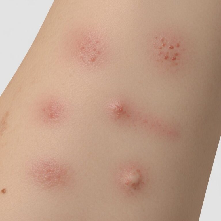
Arthropod reactions, including insect bites and other arthropod-related dermatitis, can present as cellulitis mimics. The presentation may vary from numerous urticarial papules near the site of a bite to generalized, large, indurated, erythematous plaques reminiscent of cellulitis. Lesions usually develop within hours of a bite and persist for an average of 1 to 2 weeks. The areas typically affected are the head and neck or the upper or lower extremities; the palms, soles, and trunk are usually spared. A careful history of recent arthropod exposure and the temporal relationship between bite and lesion development helps confirm the diagnosis.
Capillaritis
Capillaritis may be unilateral or bilateral, acute, chronic, or recurrent and favors the lower legs. It is characterized by “cayenne-pepper” petechiae, but these may resemble cellulitis when confluent and accompanied by erythematous patches and edema. Key distinguishing features include a cool leg with no systemic symptoms. The absence of warmth and fever helps differentiate this condition from true cellulitis.
Lymphoedema and Associated Conditions
Lymphoedema is associated with a risk of cutaneous infection, including cellulitis, due to impaired lymphatic drainage. However, lymphoedema itself may present with erythema and edema that can be mistaken for cellulitis. Lymphoedema typically develops gradually and may be preceded by cancer treatment, surgery, or congenital lymphatic abnormalities. The absence of acute systemic symptoms and the chronic nature of the presentation help differentiate lymphoedema from acute cellulitis.
Diagnostic Approach and Differentiation
Given the high rate of cellulitis misdiagnosis, a systematic approach to differential diagnosis is essential. A symmetric or diffusely scattered pattern indicates a condition other than cellulitis, which is overwhelmingly unilateral with smooth, indistinct borders. Other factors pointing to cellulitis are underlying immunosuppression, more rapid progression, previous episodes, and systemic symptoms such as fever and malaise.
The dermatologist can determine if biopsy is necessary, as many dermatoses that mimic cellulitis can be diagnosed by visual recognition alone. For uncertain cases, dermatology consultation is recommended to narrow the differential diagnosis. Imaging studies such as ultrasound, CT, or MRI scans can be helpful in assessing deeper tissue involvement, particularly when distinguishing cellulitis from necrotising fasciitis or deep abscesses.
Key Diagnostic Clues Summary
| Condition | Key Distinguishing Features | Distribution |
|---|---|---|
| Cellulitis | Unilateral, warm, tender, indistinct borders, systemic symptoms, rapid onset | Unilateral |
| Stasis Dermatitis | Bilateral, cool, bound-down plaques, chronic history, no fever | Bilateral lower legs |
| Contact Dermatitis | History of exposure, vesicles, scaling, pruritus, confined to contact area | Localized or symmetric |
| Erysipelas | Sharply demarcated raised borders, orange peel texture, facial involvement common | Often facial or lower extremities |
| Eosinophilic Cellulitis | Recurrent lesions, eosinophils on biopsy, fading over weeks | Multiple lesions |
| Herpes Zoster | Dermatomal distribution, umbilicated vesicles, antiviral responsive | Dermatomal |
Avoiding Unnecessary Antibiotic Use
Accurate differentiation between cellulitis and its mimics is crucial for avoiding unnecessary antibiotic exposure, which contributes to the development of antibiotic-resistant bacteria. Many conditions that mimic cellulitis require entirely different treatment approaches. For example, contact dermatitis requires removal of the offending agent and supportive care, while eosinophilic cellulitis may require immunosuppressive therapy. Herpes zoster requires antiviral agents, not antibiotics. By correctly identifying these conditions, healthcare providers can tailor treatment appropriately and prevent adverse outcomes associated with unnecessary antimicrobial therapy.
Frequently Asked Questions
Q: What percentage of cellulitis diagnoses are actually misdiagnoses?
A: Approximately 30% of cases diagnosed as cellulitis are misdiagnosed, meaning the patient actually has a different condition mimicking cellulitis. More than 10% of patients labeled as having cellulitis do not have cellulitis.
Q: How can I distinguish cellulitis from stasis dermatitis?
A: The most important distinguishing feature is that cellulitis is unilateral while stasis dermatitis is typically bilateral. Cellulitis presents with warmth and tenderness with rapid acute onset, while stasis dermatitis has a chronic progression, cool skin, and bound-down plaques. Stasis dermatitis does not cause systemic symptoms like fever.
Q: What clinical clues suggest a diagnosis other than cellulitis?
A: A symmetric or diffusely scattered pattern indicates a condition other than cellulitis. Additional clues include absence of systemic symptoms, chronic rather than acute presentation, cool rather than warm skin, vesicles or scaling, history of allergen exposure, or dermatomal distribution suggesting herpes zoster.
Q: When should I refer a patient with suspected cellulitis to a dermatologist?
A: Referral is recommended when the diagnosis is uncertain, when features are atypical for cellulitis, when the condition fails to respond to appropriate antibiotics, or when the presentation is bilateral or symmetric. The dermatologist can determine if skin biopsy is necessary, as many dermatoses mimicking cellulitis can be diagnosed by visual recognition alone.
Q: Can contact dermatitis be secondarily infected with bacteria?
A: Yes, contact dermatitis can become secondarily infected, complicating the clinical picture and making it appear more like cellulitis. However, the history of allergen exposure and the pattern of distribution confined to the contact area help differentiate it from true cellulitis.
Q: What distinguishes necrotising fasciitis from cellulitis?
A: Necrotising fasciitis presents with severe pain disproportionate to clinical findings, rapid progression over hours, systemic toxicity, and skin changes including erythema, bullae, and necrosis. It is a medical emergency requiring immediate surgical intervention, whereas uncomplicated cellulitis responds to antibiotics alone.
References
- Distinguishing cellulitis from its mimics — Kenneth J. Tomecki, MD, Cleveland Clinic Journal of Medicine. 2012-08. https://www.ccjm.org/content/ccjom/79/8/547.full.pdf
- Cellulitis mimics — DermNet New Zealand. 2025. https://dermnetnz.org/topics/cellulitis-mimics
- Misdiagnosis of Uncomplicated Cellulitis: a Systematic Review and Meta-Analysis — National Center for Biotechnology Information, U.S. National Library of Medicine. 2023. https://pmc.ncbi.nlm.nih.gov/articles/PMC10406744/
- Skin Infections In People Who Inject Drugs — DermNet New Zealand. 2025. https://dermnetnz.org/topics/skin-infections-in-people-who-inject-recreational-drugs
- Cellulitis — StatPearls, National Center for Biotechnology Information, U.S. National Library of Medicine. 2024. https://www.ncbi.nlm.nih.gov/books/NBK549770/
Similar Articles


Read full bio of medha deb