Cerebral Arteriogram: Procedure, Purpose & Risks
Complete guide to cerebral arteriograms: understanding this diagnostic imaging procedure for brain blood vessels.

What is a Cerebral Arteriogram?
A cerebral arteriogram, also known as a cerebral angiogram or cerebral angiography, is a minimally invasive diagnostic imaging procedure that uses specialized X-ray technology to visualize the blood vessels in the brain. This procedure combines catheter-based intervention with real-time radiographic imaging to create detailed pictures of arterial and venous blood flow within the cranial vault. Unlike standard X-ray imaging, which provides limited information about soft tissue structures, a cerebral arteriogram employs contrast material (a special dye) injected directly into cerebral blood vessels to make them visible on radiographic images.
The procedure represents a significant advancement in neuroimaging technology, allowing clinicians to obtain high-resolution, three-dimensional reconstructions of cerebral vasculature. This level of detail is essential for diagnosing complex vascular pathologies and planning surgical interventions. The arteriogram has become the gold standard for visualizing fine vascular anatomy and detecting subtle abnormalities that may not be apparent on other imaging modalities.
Why Your Healthcare Provider May Recommend a Cerebral Arteriogram
Physicians order cerebral arteriograms for various diagnostic and therapeutic purposes. This procedure is particularly valuable when other imaging modalities have provided inconclusive results or when definitive vascular anatomy needs to be established before surgical planning. The following represents the primary clinical indications:
- Aneurysm Detection and Evaluation: Identifying and characterizing ballooning of cerebral arteries that may rupture and cause hemorrhagic stroke
- Arteriovenous Malformations (AVMs): Detecting abnormal connections between arteries and veins in the brain
- Atherosclerotic Disease: Identifying narrowing of cerebral arteries caused by plaque buildup
- Vasculitis Evaluation: Assessing inflammation of blood vessels that causes arterial narrowing
- Vasospasm Assessment: Detecting blood vessel spasms causing irregular narrowing, particularly following subarachnoid hemorrhage
- Thrombosis and Occlusion: Locating blood clots within cerebral vessels or complete blockages of arterial flow
- Vascular Clip Evaluation: Assessing previously placed surgical clips on cerebral blood vessels
- Stroke Evaluation: Identifying the vascular source of ischemic stroke and evaluating collateral circulation
Your healthcare provider may have additional reasons specific to your clinical situation for recommending this procedure. It is essential to discuss the specific indication with your physician to understand how the results will inform your treatment plan.
How the Cerebral Arteriogram Procedure Works
Pre-Procedure Preparation
Before your cerebral arteriogram, you will receive specific instructions regarding fasting, medication management, and what to expect. You should inform your healthcare team of any allergies, particularly to iodine or contrast materials, as the contrast dye used in this procedure contains iodine. Additionally, notify your provider of any bleeding disorders, current anticoagulant medications, or renal insufficiency, as these factors may affect procedural planning.
On the day of the procedure, you will be asked to change into a hospital gown and remove all metal objects, including jewelry, watches, and hearing aids, as these can interfere with radiographic imaging. An intravenous (IV) line will be placed to administer sedative medications and contrast material. Your bladder should be emptied before the procedure begins, as the examination can take up to three hours.
Positioning and Monitoring
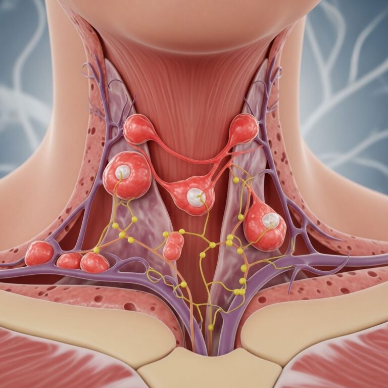
Once you are positioned on the X-ray table, your head will be secured using straps, tape, or sandbags to prevent movement during imaging. An electrocardiogram (ECG) monitor will be attached using sticky patches called leads placed on your arms and legs to continuously record your heart’s electrical activity. Your heart rate, blood pressure, breathing rate, and oxygen saturation will be monitored throughout the entire procedure by trained medical personnel.
If the catheter will be inserted in your groin or arm, the radiologist will examine the pulses below the insertion site and mark them with a marker pen. This marking allows for post-procedure verification that circulation to the limb has not been compromised.
Catheter Insertion and Navigation
You will receive mild sedation to help you relax during the procedure, though you will remain conscious and able to communicate with the medical team. The skin over the insertion site (typically the groin, sometimes the wrist, and rarely the neck) will be thoroughly cleaned with an antiseptic solution. Local anesthetic medication will be injected to numb the area, and you may feel a slight burning or stinging sensation as the anesthetic takes effect.
A small incision, typically 1/8 inch in diameter, is made in the skin over the target artery. The radiologist then inserts a thin, flexible plastic tube called a catheter—which is considerably smaller than a pencil lead—through this tiny opening into the arterial system. You may experience a sensation of pressure as the catheter is being advanced, but this should not be painful due to the local anesthetic.
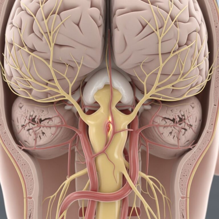
Using real-time X-ray imaging called fluoroscopy, the radiologist carefully threads the catheter through the arterial system toward the blood vessels of the brain. Fluoroscopy provides continuous imaging guidance, allowing the physician to navigate the catheter precisely to the target vessels. The entire advancement is performed without pain, though you may feel pressure sensations as the catheter moves through the vessels.
Contrast Injection and Image Acquisition
Once the catheter is correctly positioned within the cerebral vasculature, contrast material is injected through the catheter into the blood vessels. A special machine called a power injector delivers the contrast material at a precise rate and volume to ensure optimal image quality. The contrast dye, which contains iodine, temporarily highlights the blood vessels, making them visible on X-ray images.
As the contrast material flows through the cerebral circulation, multiple sets of high-resolution X-ray images are obtained from various angles. The first series typically shows arterial phase images, followed by capillary phase images, and finally venous phase images as the contrast material moves through different components of the cerebral circulation. This sequential imaging provides comprehensive information about blood flow dynamics and vascular morphology.
Advanced imaging techniques, such as digital subtraction angiography (DSA), may be employed during your procedure. With DSA, a computer automatically removes bone and tissue from the images, displaying only the blood vessels filled with contrast material. This technique significantly enhances visualization of vascular structures and abnormalities. Additionally, the acquired images can be processed to generate three-dimensional reconstructions of the cerebral vasculature, which are invaluable for surgical planning and understanding complex vascular anatomy.
Depending on the clinical indication, your radiologist may inject contrast material multiple times to examine different arterial systems or to obtain additional images from different projections.
Post-Procedure Closure and Recovery
Once all necessary images have been obtained, the radiologist removes the catheter from the artery. Pressure is immediately applied over the puncture site to prevent bleeding and hematoma formation. This manual compression is typically maintained for approximately 10 minutes. Alternatively, the radiologist may use a vascular closure device—a small plug inserted as the catheter is withdrawn—which rapidly seals the puncture site in the arterial wall. The use of closure devices allows you to resume normal activity more quickly, as the puncture site is sealed immediately rather than requiring prolonged bed rest.
After the bleeding has stopped, a sterile dressing is applied over the puncture site. A sandbag or other heavy item may be placed over the site to provide additional pressure and prevent further bleeding or hematoma formation.
What to Expect During Recovery
Following your cerebral arteriogram, you will be monitored in a recovery area for a period of time determined by your physician. During recovery, medical staff will assess your neurological status, checking for any signs of complications such as weakness, difficulty speaking, vision changes, or altered consciousness. Your puncture site will be inspected regularly for signs of bleeding or hematoma formation.
Most patients experience only mild discomfort at the puncture site following the procedure. You may notice slight bruising or swelling at the insertion site, which is normal and typically resolves within a few days. If a manual compression technique was used, you may need to remain relatively still for several hours to allow the puncture site to seal completely. If a vascular closure device was used, you may be able to move about more quickly.
Your healthcare provider will give you specific post-procedure instructions regarding activity restrictions, wound care, and when you can resume normal activities. Most patients can return to regular activities within one to two weeks, though strenuous exercise should be avoided for a longer period. Ensure that the puncture site remains clean and dry, and watch for signs of infection such as increased redness, warmth, drainage, or fever.
Risks and Complications
Like all medical procedures, cerebral arteriography carries certain risks, though serious complications are uncommon when performed by experienced interventional radiologists. Potential complications include:
- Arterial Puncture Site Complications: Bleeding, bruising, hematoma formation, or infection at the catheter insertion site
- Contrast Reaction: Allergic reactions to the iodine-containing contrast material, ranging from mild rash to severe anaphylaxis
- Thromboembolic Events: Blood clot formation at the catheter tip or within the artery, potentially leading to stroke
- Dissection: Tearing of the arterial wall during catheter manipulation
- Vasospasm: Temporary constriction of cerebral blood vessels in response to catheter manipulation
- Contrast-Induced Nephropathy: Reduced kidney function following contrast material injection, particularly in patients with pre-existing renal disease
- Neurological Complications: Transient ischemic events or stroke due to catheter manipulation or thromboembolic phenomena
Your radiologist will discuss these risks with you before the procedure and will take measures to minimize the likelihood of complications.
Important Considerations and Precautions
Allergies and Medical History
It is crucial to inform your healthcare provider of any known allergies, particularly to iodine, shellfish, or previous reactions to contrast materials. Additionally, report any medical conditions, including kidney disease, diabetes, heart disease, or bleeding disorders, as these may affect your candidacy for the procedure or require special precautions.
Medications
Inform your physician of all medications you are taking, especially anticoagulants (blood thinners) such as warfarin, apixaban, or rivaroxaban, as well as antiplatelet agents like aspirin or clopidogrel. Depending on your clinical situation, your provider may ask you to discontinue certain medications before the procedure or to continue them with special precautions.
Hydration and Contrast Material
Adequate hydration before and after the procedure helps reduce the risk of contrast-induced nephropathy, a temporary reduction in kidney function that can occur following contrast material injection. Your physician may recommend increased fluid intake in the days before your procedure and will likely encourage you to drink plenty of fluids following the arteriogram.
Frequently Asked Questions
Q: Is a cerebral arteriogram painful?
A: The procedure should not be painful. Local anesthetic is administered at the puncture site, and you will receive mild sedation to help you relax. You may experience pressure sensations as the catheter is advanced, but these should not be painful. If you experience significant pain during the procedure, inform the medical team immediately.
Q: How long does a cerebral arteriogram take?
A: The procedure typically takes one to three hours, depending on the complexity of the vascular anatomy being examined and whether therapeutic interventions are performed. You should plan to spend additional time in pre-procedure preparation and post-procedure recovery.
Q: What is the difference between a cerebral arteriogram and other brain imaging tests?
A: A cerebral arteriogram provides more detailed visualization of cerebral blood vessels than non-invasive imaging tests such as CT or MRI angiography. It is the gold standard for vascular imaging and allows for both diagnosis and treatment in a single procedure. However, it does carry slightly higher risks than non-invasive tests due to the catheter insertion.
Q: Can I eat or drink before my cerebral arteriogram?
A: You will typically be asked to fast for a specific period before the procedure (usually 6-8 hours). Your physician will provide specific fasting instructions as part of your pre-procedure preparation.
Q: What happens if an abnormality is found during my arteriogram?
A: Depending on the abnormality and your clinical situation, your interventional radiologist may perform therapeutic intervention during the same procedure, such as coil embolization for an aneurysm or stent placement for a stenotic artery. In other cases, your physician will discuss treatment options with you based on the findings.
Q: Is radiation exposure from a cerebral arteriogram dangerous?
A: The radiation dose from a cerebral arteriogram is higher than from a standard X-ray but is generally considered acceptable for the diagnostic or therapeutic benefit obtained. Your interventional radiologist uses techniques to minimize radiation exposure while maintaining image quality.
Q: When will I receive the results of my cerebral arteriogram?
A: Your radiologist will review the images immediately after the procedure. A preliminary report is typically available within hours, with a comprehensive written report usually available within 24 hours. Your referring physician will discuss the results with you and explain what they mean for your care.
References
- Cerebral Angiogram Procedure: Unveiling the Power of this Test — New Jersey Brain and Spine. 2024. https://njbrainspine.com/power-of-cerebral-angiogram/
- Cerebral Angiogram — University of Rochester Medical Center. 2024. https://www.urmc.rochester.edu/encyclopedia/Content?contentTypeID=92&ContentID=P07665
- Cerebral Angiography — Radiological Society of North America. 2024. https://www.radiologyinfo.org/en/info/angiocerebral
- Cerebral Angiography Information — Mount Sinai Health System. 2024. https://www.mountsinai.org/health-library/tests/cerebral-angiography
- Brain Angiogram: Before Your Procedure — Alberta Health Services. 2024. https://myhealth.alberta.ca/Health/aftercareinformation/pages/conditions.aspx?hwid=abr5930
Similar Articles


Read full bio of medha deb