Cerebrospinal Fluid (CSF) Leak: Causes, Symptoms & Treatment
Complete guide to CSF leaks: understand causes, recognize symptoms, and explore treatment options.
 Leak: Causes, Symptoms & Treatment")
Understanding Cerebrospinal Fluid (CSF) Leak
Cerebrospinal fluid is a vital fluid that bathes and protects the brain and spinal cord, acting as a cushion against trauma and injury. A cerebrospinal fluid (CSF) leak occurs when there is an abnormal opening or communication between the membrane surrounding the brain and spinal cord (the dura mater) and the spaces outside this membrane. This leakage allows CSF to escape from its normal compartment, leading to a range of symptoms and potential complications. CSF leaks can occur in different locations and for various reasons, each requiring specific diagnostic and treatment approaches.
Types of CSF Leaks
Understanding the type of CSF leak is crucial for determining appropriate treatment and managing symptoms effectively. There are two primary categories of cerebrospinal fluid leaks, each with distinct characteristics and treatment considerations.
Spinal CSF Leak
A spinal cerebrospinal fluid leak occurs anywhere along the spinal column, typically affecting the membrane surrounding the spinal cord. The most common symptom associated with spinal CSF leaks is a positional headache that significantly worsens when standing upright and improves when lying down. This characteristic positional nature helps distinguish spinal leaks from other types of headaches. Patients with spinal CSF leaks often experience nausea, vomiting, and a sensation of heaviness or stiffness in the neck and shoulders. Many spinal CSF leaks can be managed conservatively with bed rest, increased fluid intake, and anti-inflammatory medications, though some may require more intensive intervention.
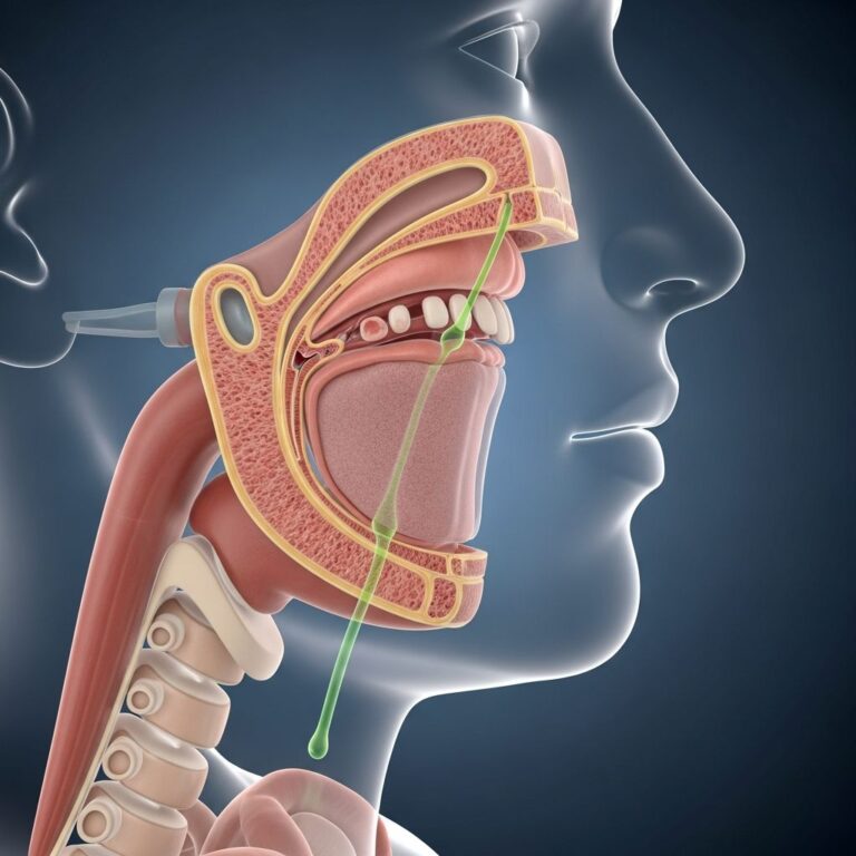
Cranial CSF Leak
A cranial cerebrospinal fluid leak occurs in the skull or cranium, typically involving a communication between the brain’s protective lining and the nasal cavity or ear canal. Unlike spinal leaks, cranial CSF leaks often produce a distinctive symptom: watery, salty discharge from the nose (rhinorrhea) or ear (otorrhea), usually from one side of the body. These leaks frequently cause chronic headaches and may be associated with hearing loss and neck pain. The location and severity of cranial CSF leaks often determine whether conservative management or surgical intervention is necessary.
Causes of CSF Leaks
Cerebrospinal fluid leaks can develop through various mechanisms, ranging from traumatic injuries to medical procedures and structural abnormalities. Understanding the underlying cause is essential for determining the most appropriate treatment strategy.
Common Causes of Spinal CSF Leaks
Spinal CSF leaks can result from multiple factors, including:
- Lumbar puncture or spinal tap procedures, which are diagnostic tests that involve inserting a needle into the spinal canal
- Epidural anesthesia administered for pain relief during procedures or labor and delivery
- Head or spine injuries and trauma
- Bone spurs or calcified disc herniations that damage the dura mater
- Congenital birth defects affecting the spinal structures
- Dura mater and nerve root abnormalities
- Irregular connections between the dura mater and veins, known as CSF-venous fistulas
- Prior spine surgery
- Repetitive activities such as heavy lifting, strenuous exercise, or sudden falls
- Forceful activities like coughing, sneezing, or straining during bowel movements
Causes of Cranial CSF Leaks
Cranial cerebrospinal fluid leaks are most commonly caused by trauma to the head or face. Additionally, prior nasal or sinus surgery can create openings that allow cerebrospinal fluid to leak into the nasal cavity. Some cranial CSF leaks develop spontaneously when patients have elevated intracranial pressure, similar to high blood pressure affecting the systemic circulation. In these cases, the elevated pressure forces cerebrospinal fluid through small openings between the brain and nasal cavity. In some instances, both cranial and spinal CSF leaks may not have an identifiable cause, classified as spontaneous or idiopathic leaks.
Symptoms and Clinical Presentation
The symptoms of a CSF leak vary significantly depending on whether the leak is spinal or cranial in location. Recognizing these symptoms is crucial for seeking appropriate medical evaluation and treatment.
Spinal CSF Leak Symptoms
Patients with spinal cerebrospinal fluid leaks typically experience:
- Positional headaches that worsen when standing and improve when lying flat
- Nausea and vomiting
- Imbalance or dizziness, particularly upon standing
- Neck and shoulder pain, numbness, or stiffness
- Tinnitus or ringing in the ears
- Hearing changes or loss
- Visual disturbances or blurred vision
- Sensitivity to light and sound
- Brain fog, difficulty concentrating, and cognitive difficulties
- Photophobia
Cranial CSF Leak Symptoms
Patients with cranial cerebrospinal fluid leaks typically present with:
- Clear, watery, salty-tasting fluid draining from the nose or ear, often from one side
- Chronic headaches
- Hearing loss or changes in hearing
- Neck pain and stiffness
- Positive halo sign (clear fluid that stains a ring on gauze due to glucose content)
Diagnostic Procedures
Accurate diagnosis of a cerebrospinal fluid leak requires multiple diagnostic approaches to confirm the presence of CSF leakage and pinpoint its exact location. A combination of laboratory tests and imaging studies provides the most comprehensive assessment.
Laboratory Testing
The first diagnostic step involves collecting a small sample of the fluid suspected to be cerebrospinal fluid and testing it for beta-2 transferrin, a protein found exclusively in cerebrospinal fluid. This laboratory confirmation definitively establishes whether the discharged fluid is indeed CSF, helping distinguish it from other fluids such as mucus or serous discharge.
Imaging Studies
Once CSF leakage is confirmed, various imaging techniques help locate the exact site of the leak and determine the most appropriate treatment strategy.
Myelography: This imaging test combines a contrast dye with X-rays or computed tomography (CT) to create detailed pictures of the spinal column. Myelography can identify the precise location of a spinal CSF leak and helps determine the most appropriate treatment plan. A spinal tap is typically required before myelography to introduce the contrast agent into the cerebrospinal fluid.
MRI with Gadolinium: Magnetic resonance imaging using gadolinium contrast is particularly useful for detecting cerebrospinal fluid leaks within the brain itself. Gadolinium, a contrast agent, highlights irregularities in brain tissue and helps locate the source of cranial CSF leaks.
CT/MRI Fusion Technique: Advanced imaging centers employ fusion techniques that combine CT and MRI images to provide superior visualization of leak locations, particularly for complex cranial leaks. This multimodal approach allows neurosurgeons to plan surgical interventions with greater precision.
Specialized Diagnostic Tests
Spinal Tap (Lumbar Puncture): This procedure involves inserting a needle into the spinal canal to measure the pressure of cerebrospinal fluid. It is often performed as part of the diagnostic workup for suspected CSF leaks and is typically required as a preliminary step before myelography or cisternography.
Tympanometry: This test uses a handheld device called a tympanometer, which is inserted into the ear canal to measure middle ear function and check for fluid accumulation. It is particularly useful when evaluating potential cerebrospinal fluid leaks that may present as fluid in the middle ear.
Treatment Options
Treatment approaches for cerebrospinal fluid leaks range from conservative management strategies to minimally invasive procedures and surgical repair. The choice of treatment depends on the leak’s location, severity, underlying cause, and patient symptoms.
Conservative Treatment Approaches
Many cerebrospinal fluid leaks, particularly those caused by trauma or recent medical procedures, can improve with conservative management strategies that reduce symptoms and allow spontaneous healing.
Symptom Management: Conservative approaches involve managing symptoms as they develop through several methods. Bed rest and horizontal positioning help reduce cerebrospinal fluid pressure gradients, minimizing leakage. Oral and intravenous hydration provide temporary symptomatic relief by replenishing cerebrospinal fluid volume. Oral and intravenous caffeine can temporarily improve symptoms by affecting fluid dynamics. Additionally, stool softeners prevent straining that could exacerbate leakage.
Complementary Approaches: Some patients benefit from complementary therapies including nutritional support, dietary supplements, mind-body techniques, and acupuncture, though evidence supporting these interventions varies.
Epidural Blood Patch (EBP)
The epidural blood patch is a minimally invasive procedure that has proven effective for many patients with cerebrospinal fluid leaks. During this procedure, a small sample of the patient’s own blood is extracted and then injected into the epidural space, which is the area just outside the dura mater within the spinal canal. The injected blood cells form a clot, which creates a biological “patch” over the area where cerebrospinal fluid is leaking, effectively sealing the defect. This procedure is typically performed by anesthesiologists or radiologists using fluoroscopic guidance and intravenous sedation to ensure accuracy and patient comfort. A substantial percentage of patients respond favorably to one or more epidural blood patching procedures, making it a first-line intervention for many spinal CSF leaks.
Fibrin Sealant Injection
Fibrin sealant is a special biological glue composed of substances derived from human plasma that promote blood clotting. This sealant can be injected alone or mixed with the patient’s own blood and delivered into the spinal canal to cover the hole and stop cerebrospinal fluid leakage. This procedure is most often performed by neuroradiologists using imaging guidance and intravenous sedation to target specific known or suspected leak locations. It may be used in isolation or in combination with whole blood for enhanced effectiveness.
Trans-Venous Embolization
This minimally invasive procedure is specifically designed for CSF-venous fistulas, which are abnormal connections between cerebrospinal fluid spaces and blood vessels in the spine. Trans-venous embolization works by “gluing shut” the fistula from inside the affected vein, effectively stopping the cerebrospinal fluid leak into blood vessels and preventing associated complications.
Surgical Repair
When conservative measures and minimally invasive procedures fail or when the leak’s location is unsuitable for patching procedures, surgical repair becomes necessary. Surgical interventions are particularly important for cranial cerebrospinal fluid leaks that require closure of the communication between the brain and nasal cavity. During surgical repair, tissue from within the nasal passages is used to seal the defect, and adjacent sinuses are opened to prevent future chronic sinusitis. Patients undergoing surgical repair are typically discharged home within one to two days and receive close office follow-up to ensure proper healing of the nasal cavity. Surgical repairs performed by experienced neurosurgeons demonstrate good success rates, though some patients may experience persistent symptoms requiring additional intervention.
Adjunctive Treatment for Elevated Pressure
Patients with spontaneous cerebrospinal fluid leaks caused by elevated intracranial pressure may require additional procedures such as a shunt to control the problematic pressure elevation around the brain. This adjunctive treatment addresses the underlying pressure issue to prevent future cerebrospinal fluid leaks and complications related to increased intracranial pressure, such as vision and hearing problems.
Complications and Risks
Untreated or inadequately managed cerebrospinal fluid leaks can lead to serious complications. Persistent leakage results in intracranial hypotension, causing headache, confusion, nausea, dizziness, and vomiting. Subdural hematoma, characterized by bleeding in the space between the brain and skull, can cause brain sagging, tear bridging veins, and create pressure on brain tissue. If not identified promptly, this condition constitutes a surgical emergency requiring urgent intervention.
Recovery and Follow-Up Care
Recovery from cerebrospinal fluid leak treatment varies depending on the approach used. Patients undergoing conservative management require consistent bed rest and follow-up evaluations to monitor symptoms. Those receiving epidural blood patches or fibrin sealant injections typically experience symptom improvement within hours to days. Surgical patients require closer monitoring during the initial healing phase, with most returning to normal activities within several weeks. Regular follow-up appointments with healthcare providers ensure proper healing and allow for early identification of any recurrent symptoms or complications.
Frequently Asked Questions About CSF Leaks
Q: Can a cerebrospinal fluid leak heal on its own?
A: Yes, many cerebrospinal fluid leaks, particularly those caused by procedures like lumbar puncture or epidural anesthesia, can heal spontaneously with conservative treatment including bed rest, hydration, and anti-inflammatory medications. However, some leaks require medical intervention to ensure proper healing and prevent complications.
Q: How long does it take for a CSF leak to heal?
A: The healing timeline varies depending on the cause and severity of the leak. Conservative treatment may take days to weeks, while epidural blood patches typically provide relief within 24-48 hours. Surgical repairs generally allow patients to resume normal activities within several weeks.
Q: Is epidural blood patch safe?
A: Yes, epidural blood patch is generally considered a safe procedure with high success rates. It uses the patient’s own blood, minimizing rejection risks, and is performed by experienced healthcare professionals using imaging guidance to ensure proper placement.
Q: Can a cerebrospinal fluid leak recur after treatment?
A: While most patients experience successful resolution with appropriate treatment, some may experience symptom recurrence. If initial treatments are unsuccessful, physicians can guide further intervention options, including additional patches or surgical repair.
Q: What should I do if I suspect I have a cerebrospinal fluid leak?
A: Seek medical attention immediately if you experience symptoms such as positional headaches, clear nasal or ear discharge, or neurological symptoms. Early diagnosis and appropriate treatment can prevent complications and improve outcomes.
References
- CSF leak (Cerebrospinal fluid leak) – Diagnosis and treatment — Mayo Clinic. 2024. https://www.mayoclinic.org/diseases-conditions/csf-leak/diagnosis-treatment/drc-20522247
- CSF leak (Cerebrospinal fluid leak) – Symptoms and causes — Mayo Clinic. 2024. https://www.mayoclinic.org/diseases-conditions/csf-leak/symptoms-causes/syc-20522246
- Cerebrospinal Fluid (CSF) Leak: Symptoms & Causes — NewYork-Presbyterian. 2024. https://www.nyp.org/ochspine/cerebrospinal-fluid-csf-leak
- Cerebrospinal Fluid Leak – StatPearls — National Center for Biotechnology Information (NCBI) Bookshelf. 2024. https://www.ncbi.nlm.nih.gov/books/NBK538157/
- Cerebrospinal Fluid (CSF) Leak Symptoms and Treatment NYC — Mount Sinai Health System. 2024. https://www.mountsinai.org/locations/skull-base-surgery-center/conditions/cerebrospinal-fluid-leak
- Treatment – Spinal CSF Leak Foundation — Spinal CSF Leak Foundation. 2024. https://spinalcsfleak.org/about-spinal-csf-leaks/treatment/
- Cerebrospinal Fluid Leak — Cedars-Sinai. 2024. https://www.cedars-sinai.org/health-library/diseases-and-conditions/c/cerebrospinal-fluid-leak.html
Similar Articles


Read full bio of medha deb