Chédiak-Higashi Syndrome: Symptoms, Diagnosis & Treatment
Rare childhood disorder causing immune dysfunction, albinism, infections, bleeding, and neurological issues.

Author: Dr. Amanda Oakley, Dermatologist, Hamilton, New Zealand. Reviewed: 25 February 2025
Introduction
Chédiak-Higashi syndrome (CHS) is a rare, autosomal recessive disorder primarily affecting children, characterized by severe immune dysfunction, partial oculocutaneous albinism, neurological abnormalities, and a high risk of life-threatening complications. First described in 1943 by Argentine paediatrician Arturo Béguez César, it is also known as Béguez-César syndrome or leukocyte anomaly albinism. The condition arises from mutations in the LYST gene (lysosomal trafficking regulator), leading to defective lysosomal granule formation and function in multiple cell types, including melanocytes, neutrophils, and natural killer cells. This results in hypopigmentation, recurrent pyogenic infections, easy bruising, and progressive neuropathy. Approximately 80% of untreated patients progress to an accelerated phase resembling haemophagocytic lymphohistiocytosis (HLH), which is often fatal in childhood without intervention. CHS highlights the critical role of lysosomal trafficking in immune surveillance, pigmentation, and cellular homeostasis, with hematopoietic stem cell transplantation (HSCT) remaining the only curative option for the immunological defects.
Demographics
CHS is exceedingly rare, with an estimated worldwide incidence of less than 1 in 1,000,000 live births, though exact prevalence is unknown due to underdiagnosis in resource-limited settings. It affects all ethnic groups but shows higher rates in populations with consanguinity, such as certain isolated communities or regions with cultural practices of cousin marriages. No sex predilection exists, as it follows autosomal recessive inheritance—both parents must be carriers of pathogenic LYST variants. Over 100 mutations have been identified in the LYST gene on chromosome 1q42.1-42.2, with nonsense, frameshift, and missense variants leading to absent or dysfunctional LYST protein. Symptoms typically manifest in infancy or early childhood (by age 5), though rare adult-onset forms exist with milder phenotypes, primarily neurological. Global case reports span diverse ancestries, underscoring its pan-ethnic occurrence despite genetic bottlenecks in some kindreds. Early recognition is crucial, as median survival without HSCT is under 10 years.
Causes
CHS results from biallelic pathogenic variants in the LYST (lysosomal trafficking regulator) gene, encoding a 380-kDa protein essential for vesicular trafficking, lysosome-related organelle biogenesis, and autophagosome-lysosome fusion. The LYST protein regulates the size, structure, and fission of lysosomes and related organelles (e.g., melanosomes, azurophilic granules, platelet-dense granules), preventing their abnormal enlargement. Mutations disrupt microtubule-based organelle transport, impairing phagocytosis, degranulation, chemotaxis, and cytotoxicity in leukocytes and NK cells. In melanocytes, defective melanosome maturation causes hypopigmentation; in platelets, abnormal dense granule function leads to bleeding diathesis. Pathogenic variants are inherited autosomal recessively; carrier parents are asymptomatic. Compound heterozygosity is common, with over 120 reported alleles including truncating (most severe) and hypomorphic variants (partial function, adult-onset). Prenatal diagnosis via genetic testing is feasible in at-risk families. Environmental triggers like viral infections (e.g., EBV) can precipitate the accelerated HLH phase by hyperactivating defective immune cells.
Clinical features
Clinical manifestations of CHS span multiple systems, appearing shortly after birth or by age 5, with hallmark
partial oculocutaneous albinism
due to giant, immature melanosomes impairing melanin transport. Skin and hair exhibit silvery-grey or blond sheen with hypopigmentation; eyes show photophobia, nystagmus, reduced visual acuity, and pale irides/fundi.Immunodeficiency
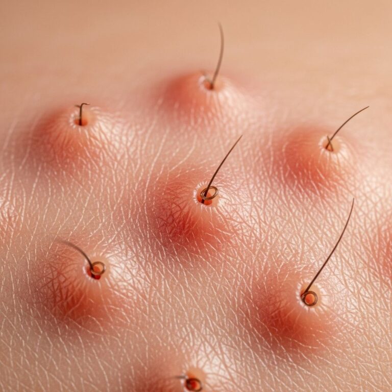
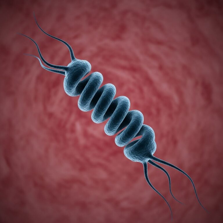
manifests as recurrent pyogenic infections (otitis media, pneumonia, skin abscesses) from neutropenia, defective chemotaxis, and impaired bactericidal activity secondary to giant azurophilic granules in granulocytes.Bleeding tendency
arises from platelet dysfunction despite normal counts, causing easy bruising, epistaxis, gingival bleeding, and prolonged post-surgical hemorrhage. Neurological features emerge later (adolescence/adulthood), including peripheral neuropathy (sensory-motor ataxia, hyporeflexia), spinocerebellar degeneration, weakness, and Parkinsonism-like tremors. Oral findings include recurrent gingivitis, periodontitis, and mucosal hypopigmentation. Adult-onset variants may present primarily with neuropathy sparing severe infections. Systemic exam reveals splenomegaly in accelerated phase.- Skin/hair: Silvery-blond hair, fair skin, photosensitivity
- Ocular: Nystagmus, photophobia, strabismus, reduced acuity
- Infections: Bacterial (Staph/Strep), viral, fungal; lung/skin/ear predominant
- Hematologic: Bruising, petechiae, prolonged bleeding time
- Neurologic: Ataxia, neuropathy, weakness (later onset)
Complications
The most dreaded complication is the
accelerated phase
(HLH-like), affecting 50–85% of patients, typically by age 4, triggered by infections (e.g., EBV, CMV). It features fever, hepatosplenomegaly, pancytopenia, hypertriglyceridemia, hypofibrinogenemia, and hemophagocytosis in bone marrow, driven by uncontrolled T-cell/NK-cell activation and cytokine storm. Untreated mortality exceeds 90% within weeks. Infections contribute to 30–50% of deaths pre-HSCT. Neurological progression causes disability, with axonal neuropathy leading to wheelchair dependence. Ocular complications include severe myopia and corneal scarring. Bleeding episodes risk intracranial hemorrhage. Post-HSCT, graft-versus-host disease and transplant-related mortality occur in 15–20%. Long-term survivors face infertility, malignancy risk, and persistent neuropathy despite immune reconstitution.Diagnosis
CHS diagnosis combines clinical suspicion with confirmatory tests.
Peripheral blood smear
reveals pathognomonicgiant azurophilic granules
(>5 μm) in neutrophils, lymphocytes, and monocytes—diagnostic in 90% of cases. Flow cytometry shows absent NK-cell cytotoxicity and reduced LAMP-1/CD107a expression.Genetic testing
confirms biallelic LYST variants via sequencing. Bone marrow biopsy in accelerated phase demonstrates hemophagocytosis. Skin biopsy shows giant melanosomes on electron microscopy. Differentiate from Griscelli syndrome (normal granules), Hermansky-Pudlak (no infections), and Vici syndrome (agenesis of corpus callosum). Prenatal diagnosis uses amniocentesis/CVS for at-risk pregnancies.| Test | Finding in CHS |
|---|---|
| Blood smear | Giant granules in leukocytes |
| Genetic | LYST mutation |
| NK function | Impaired cytotoxicity |
| Hair microscopy | Giant melanosomes |
Treatment
**Hematopoietic stem cell transplantation (HSCT)** is curative for immune defects and prevents HLH if performed pre-accelerated phase (success >80% with HLA-matched donors). For HLH, initial management includes etoposide, dexamethasone, and cyclosporine per HLH-2004 protocol, bridging to HSCT. Vitamin C (high-dose) stabilizes granules, reducing infections in non-transplanted patients. Aggressive antimicrobials (antibiotics, antifungals, acyclovir) treat/prevent infections. Platelet transfusions address bleeding; sun protection (SPF 50+, sunglasses) mitigates photophobia. Neurological symptoms lack specific therapy—supportive physiotherapy. Multidisciplinary care involves hematology, immunology, neurology, ophthalmology, and genetics.
Outlook
Without HSCT, prognosis is dismal: 50–85% enter fatal HLH by age 10, with median survival <7 years from infections/bleeding. Post-HSCT, 5-year survival exceeds 80%, curing immunodeficiency but not pre-existing neuropathy or albinism. Adult-onset forms have near-normal lifespan with milder symptoms. Ongoing research explores gene therapy targeting LYST. Genetic counseling is essential for families.
Frequently asked questions
What is Chédiak-Higashi syndrome?
A rare genetic disorder causing albinism, infections, bleeding, and HLH risk due to LYST mutations.
Is CHS curable?
HSCT cures immune aspects; neurological issues persist.
How is CHS diagnosed?
By giant leukocyte granules on blood smear and genetic confirmation.
What causes the accelerated phase?
Viral triggers induce cytokine storm in defective immune cells.
Can CHS be prevented?
Genetic counseling and prenatal testing for carriers.
References
- Chediak-Higashi syndrome: a clinical and molecular view of a rare lysosome-related disorder — Nagle DL et al. Pediatric Dermatology. 2002-07-01. https://pubmed.ncbi.nlm.nih.gov/12125812/
- Chediak Higashi Syndrome — National Organization for Rare Disorders (NORD). 2023-05-15. https://rarediseases.org/rare-diseases/chediak-higashi-syndrome/
- Chediak-Higashi syndrome — Osmosis from Elsevier. 2024-01-10. https://www.osmosis.org/learn/Chediak-Higashi_syndrome
- Chediak-Higashi syndrome — MedlinePlus, U.S. National Library of Medicine. 2025-03-20. https://medlineplus.gov/ency/article/001312.htm
- Chediak-Higashi syndrome — DermNet NZ. 2025-02-25. https://dermnetnz.org/topics/chediak-higashi-syndrome
- Chediak-Higashi Syndrome — Karim A et al. StatPearls [Internet]. NCBI Bookshelf. 2024-06-28. https://www.ncbi.nlm.nih.gov/books/NBK507881/
Similar Articles


Read full bio of medha deb