Chromoendoscopy: Advanced Dye-Based Visualization for Digestive Health
Discover how chromoendoscopy uses specialized dyes to detect precancerous lesions and abnormal tissue in the digestive tract.

What is Chromoendoscopy?
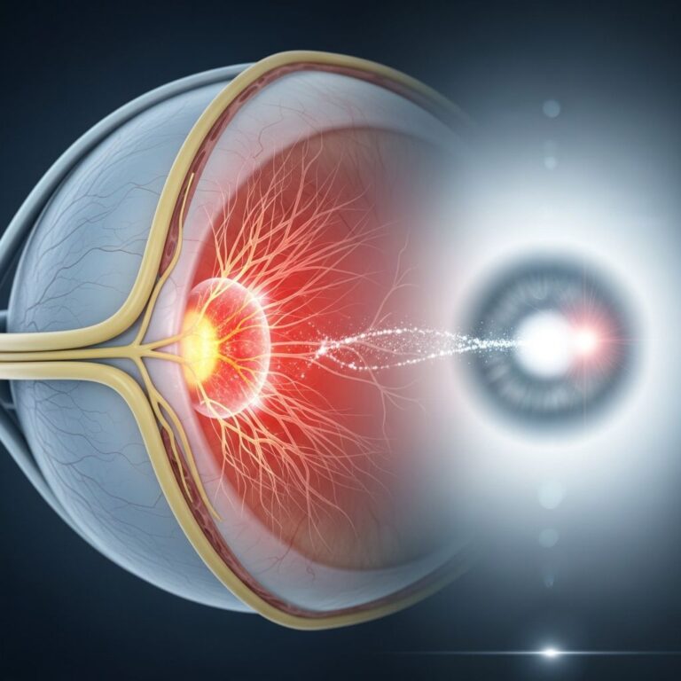
Chromoendoscopy is an advanced diagnostic technique that combines traditional endoscopy with the application of specialized dyes or stains to enhance visualization of the digestive tract. During this minimally invasive procedure, a trained gastroenterologist uses a thin, flexible tube called an endoscope equipped with a camera to examine the interior surfaces of the esophagus, stomach, and colon. The key innovation of chromoendoscopy is the application of nontoxic dye solutions that are sprayed onto the mucosal lining of the gastrointestinal tract. These dyes highlight specific areas and enhance tissue differentiation, making it significantly easier to identify abnormal tissue patterns that might otherwise be missed during conventional white-light endoscopy.
The fundamental principle behind chromoendoscopy involves using absorptive and contrast stains that interact differently with normal and abnormal tissue. Normal, healthy cells absorb these dyes uniformly, while precancerous and cancerous areas often do not pick up the dye in the same manner, creating a distinctive visual contrast. This visual enhancement allows physicians to distinguish between normal tissue and areas that require closer examination or biopsy, improving the overall diagnostic accuracy of the procedure.
How Chromoendoscopy Works
The chromoendoscopy procedure involves several carefully coordinated steps designed to maximize diagnostic accuracy. The process begins with patient preparation, which typically includes administration of sedation to ensure comfort during the examination. The endoscope, a slender instrument containing a camera and light source, is gently advanced through the mouth (for upper digestive tract examination) or rectum (for lower digestive tract examination).
Once the endoscope is properly positioned, the physician applies dye solutions through a spray catheter inserted through the working channel of the endoscope. The dye spreads across the mucosal surface, interacting with the tissue in specific ways. Normal tissue cells absorb the dye and become uniformly stained, while dysplastic (abnormal) cells typically fail to absorb the dye properly, remaining unstained or appearing with different coloration. This contrast makes abnormal areas stand out visibly to the trained endoscopist.
The procedure can be performed in two distinct approaches. Pancolonic chromoendoscopy involves applying dye throughout the entire colon on an untargeted basis, providing comprehensive screening. Alternatively, targeted chromoendoscopy directs dye application specifically to suspicious lesions or areas of concern identified during initial white-light examination. This targeted approach allows physicians to focus their diagnostic efforts on the most relevant areas, potentially reducing procedure time and improving efficiency.
Types of Dyes and Stains Used
Chromoendoscopy utilizes two primary categories of stains, each serving distinct diagnostic purposes. Absorptive or vital stains are chemical compounds that are actively taken up by cells, differentially staining normal versus abnormal tissue based on cellular characteristics. These stains penetrate the tissue and highlight specific cellular features, making them particularly useful for identifying dysplastic changes.
Non-absorptive contrast stains work through a different mechanism, simply pooling into depressions and raised areas of the mucosal surface to enhance visual definition of surface morphology. These stains do not require cellular absorption but instead highlight the physical topology and contours of the tissue surface, enhancing visualization of subtle architectural changes that may indicate pathology.
Common dyes employed in chromoendoscopy include methylene blue, which is frequently used for lower gastrointestinal tract examination; indigo carmine, a vital stain that highlights surface morphology; and cresyl violet, typically applied in small amounts (1–2 milliliters) to avoid excessive darkening of stained surfaces. For upper gastrointestinal examination, phenol red containing urea may be used, typically producing yellow to red staining within 2–3 minutes of application.
Clinical Applications and Indications
Chromoendoscopy has become an invaluable diagnostic tool for detecting and evaluating various gastrointestinal pathologies. The procedure is particularly beneficial for patients at elevated risk for colorectal cancer or those with specific digestive conditions requiring enhanced visualization.
Detection of Precancerous Lesions
One of the most important applications of chromoendoscopy is the detection of precancerous tissue growth and early-stage cancers throughout the digestive tract. The enhanced visualization provided by chromoendoscopy significantly improves the identification rate of dysplastic lesions compared to standard colonoscopy. This enhanced detection capability is especially critical for patients with a family history of colorectal cancer, personal history of adenomas, or other risk factors that increase their likelihood of developing colorectal malignancy.
Inflammatory Bowel Disease Surveillance
Patients diagnosed with inflammatory bowel disease, including ulcerative colitis and Crohn’s disease, face an elevated risk of developing colorectal cancer over time. Chromoendoscopy has become a standard surveillance tool for these populations. The enhanced visualization allows endoscopists to identify areas of dysplasia that might be invisible to standard endoscopy, enabling early intervention before cancer develops. Chromoendoscopy combined with magnifying endoscopy has significantly improved the detection of neoplastic lesions in the gastrointestinal mucosa, particularly in patients with ulcerative colitis and Crohn’s colitis.
Barrett’s Esophagus Evaluation
Barrett’s esophagus is a condition in which the normal tissue lining the esophagus is replaced by tissue resembling the stomach lining, typically as a consequence of chronic gastroesophageal reflux disease (GERD). This condition carries an increased risk of esophageal cancer development. Chromoendoscopy is particularly beneficial for observing and evaluating the esophagus in patients with Barrett’s esophagus, helping physicians identify areas of dysplasia that require targeted treatment or closer surveillance.
Other Digestive Conditions
Beyond colorectal and Barrett’s esophagus applications, chromoendoscopy helps diagnose gastric cancer (stomach cancer), esophageal cancer, and assists in the general characterization and differentiation of various endoscopically detected suspicious lesions throughout the digestive tract. The procedure aids in determining the extent of neoplastic lesions and helps identify which lesions may be amenable to endoscopic resection rather than requiring surgical intervention.
Advantages of Chromoendoscopy
Chromoendoscopy offers several significant advantages that distinguish it from conventional endoscopic procedures. The most substantial benefit is dramatically improved detection sensitivity. By highlighting areas of dysplasia that would otherwise remain invisible or appear subtle under white-light examination, chromoendoscopy enables physicians to identify precancerous and cancerous lesions at earlier, more treatable stages.
Another important advantage involves targeted tissue sampling. Rather than obtaining multiple random biopsies throughout the examined organ, physicians using chromoendoscopy can concentrate their biopsy efforts on specific areas highlighted by the dye as potentially abnormal. This targeted approach reduces the number of unnecessary biopsies, decreases histopathologic workload, improves diagnostic yield, and potentially offsets the costs associated with additional procedure time.
The procedure also provides valuable diagnostic information that might otherwise be unobtainable through conventional biopsy alone. Chromoendoscopy combined with magnifying endoscopy provides enhanced visualization of cellular architecture and can even offer accurate prediction of histology, allowing physicians to make more informed clinical decisions regarding treatment and surveillance strategies.
Preparation for Chromoendoscopy
Proper patient preparation is essential for successful chromoendoscopy. The specific preparation requirements may vary depending on which portion of the digestive tract is being examined, but general principles apply to all chromoendoscopy procedures.
For upper gastrointestinal chromoendoscopy, patients typically receive acid suppression therapy either orally the day before the procedure (using proton pump inhibitors) or intravenously 30–60 minutes before examination. Patients also receive oral anti-foaming mucolytic agents and anti-cholinergic drugs to suppress gastric motility, ensuring optimal visualization of the gastric mucosa.
For lower gastrointestinal chromoendoscopy, standard colonoscopy bowel preparation is required, including clear liquid diets and laxative agents to ensure the colon is thoroughly cleansed. Patients should follow specific fasting instructions and medication guidelines provided by their healthcare facility.
All patients undergoing chromoendoscopy receive conscious sedation administered by trained anesthesia professionals, ensuring comfort throughout the procedure while maintaining patient safety.
What to Expect During Chromoendoscopy
Understanding the procedural steps helps patients know what to anticipate during chromoendoscopy. After receiving sedation, the patient is positioned appropriately, and the endoscope is gently advanced. The physician first performs a thorough examination using standard white-light visualization, identifying any obviously abnormal areas.
The dye solution is then carefully applied to the mucosal surface using the spray catheter. The dye rapidly spreads across the examined tissue, creating visual contrast between normal and abnormal areas. The physician takes time to systematically examine all visualized areas, noting the staining pattern and any areas of concern. If abnormal tissue is identified, the physician may obtain targeted biopsies by using biopsy forceps or remove suspicious lesions using specialized instruments like snare devices. The entire procedure typically takes 15–45 minutes, depending on the extent of examination and any therapeutic interventions required.
Chromoendoscopy vs. Standard Endoscopy
While standard white-light colonoscopy remains the foundation of colorectal cancer screening, chromoendoscopy provides enhanced diagnostic capabilities for specific patient populations and clinical scenarios. Standard colonoscopy uses conventional white light to visualize the colon, which is effective for identifying larger polyps and obvious lesions but may miss subtle dysplastic changes, particularly in patients with inflammatory bowel disease or Barrett’s esophagus.
Chromoendoscopy enhances tissue differentiation through dye application, creating color highlighting that reveals surface morphology details invisible under white light. This enhanced visualization translates to superior detection rates for flat or depressed lesions and dysplastic tissue, particularly in high-risk populations. However, chromoendoscopy typically requires additional procedure time and expertise, and may not be necessary for all patients undergoing endoscopic evaluation.
Safety and Risks
Chromoendoscopy is generally a safe procedure with minimal risk when performed by experienced gastroenterologists. The dyes used are nontoxic and have been extensively studied for safety. Serious complications are rare and similar to those associated with standard colonoscopy, including rare risks of perforation (puncture of the bowel wall), bleeding (particularly if biopsies are obtained or lesions are removed), and sedation-related complications.
Mild side effects may include temporary discomfort, bloating, or mild cramping. Most patients tolerate the procedure well and experience no lasting effects. Patients should discuss any concerns or medical conditions with their healthcare provider before undergoing the procedure.
Recovery After Chromoendoscopy
Recovery from chromoendoscopy is straightforward. The sedation used during the procedure requires time to wear off, so patients should not drive or operate machinery for the remainder of the day. Most patients can resume normal activities the following day, though vigorous exercise and heavy lifting should be avoided for a few days if tissue removal or extensive biopsying was performed.
Patients may experience mild bloating or gas from air introduced during the procedure, which typically resolves within a few hours. Any severe pain, significant bleeding, or other concerning symptoms should be reported to the healthcare provider immediately.
Frequently Asked Questions
Q: How long does chromoendoscopy take?
A: The procedure typically takes 15–45 minutes depending on the extent of examination and whether biopsies or therapeutic interventions are performed. Standard preparation and recovery time add additional hours to the overall appointment duration.
Q: Is chromoendoscopy painful?
A: No, chromoendoscopy is performed under sedation, so patients remain comfortable throughout the procedure. Patients may feel slight pressure or mild cramping but should not experience pain.
Q: Can chromoendoscopy detect all types of cancer?
A: Chromoendoscopy is particularly effective for detecting precancerous and early-stage cancerous lesions in the digestive tract. However, it is most valuable for specific conditions like inflammatory bowel disease and Barrett’s esophagus. Its role in general cancer screening is still being studied.
Q: Are the dyes used in chromoendoscopy safe?
A: Yes, the dyes used in chromoendoscopy are nontoxic and have been safely used for many years. Adverse reactions are extremely rare.
Q: When will I receive my results?
A: Your physician will typically discuss preliminary findings before you leave the facility. If biopsies were obtained, definitive pathology results are usually available within 5–7 business days.
Q: Do I need chromoendoscopy if I have had a normal colonoscopy?
A: This depends on your individual risk factors and medical history. Patients at average risk typically do not need chromoendoscopy, but those with inflammatory bowel disease, Barrett’s esophagus, or strong family history of colorectal cancer may benefit from chromoendoscopy surveillance.
References
- Chromoendoscopy — Penn State Health. 2024. https://www.pennstatehealth.org/services-treatments/ibd-center/patient-care-treatment/ibd-diagnosis/chromoendoscopy
- Chromoendoscopy to Diagnose Digestive Disease — Mercy Medical Center. 2024. https://mdmercy.com/mercy-services/diagnostic-and-specialty-service/chromoendoscopy
- Indications, stains and techniques in chromoendoscopy — National Center for Biotechnology Information (NIH). 2012. https://pmc.ncbi.nlm.nih.gov/articles/PMC3550597/
- Chromoendoscopy as an Adjunct to Colonoscopy — South Carolina Blues. 2024. https://www.southcarolinablues.com/web/public/brands/medicalpolicy/external-policies/chromoendoscopy-as-an-adjunct-to-colonoscopy/
Similar Articles


Read full bio of medha deb