Chrysiasis: Comprehensive Guide To Diagnosis, Treatment & Risks
Understanding the rare gold-induced skin pigmentation: causes, clinical features, diagnosis, and management insights.

Chrysiasis is a rare dermatological condition characterized by
blue-grey to greyish-purple pigmentation
of the skin, resulting from the deposition of gold particles in the connective tissue of the dermis. This discoloration primarily affects sun-exposed areas and is a side effect of prolonged gold salt therapy, historically used for treating rheumatoid arthritis and other autoimmune diseases.What is chrysiasis?
Chrysiasis derives its name from the Greek word chrysos, meaning gold, reflecting the golden origin of the deposited particles. It manifests as permanent staining due to parenteral (intramuscular or intravenous) administration of gold salts, such as sodium aurothiomalate or aurothioglucose. The condition is iatrogenic, meaning it is physician-induced through therapeutic interventions that have largely been abandoned in modern rheumatology due to superior alternatives like methotrexate and biologics.
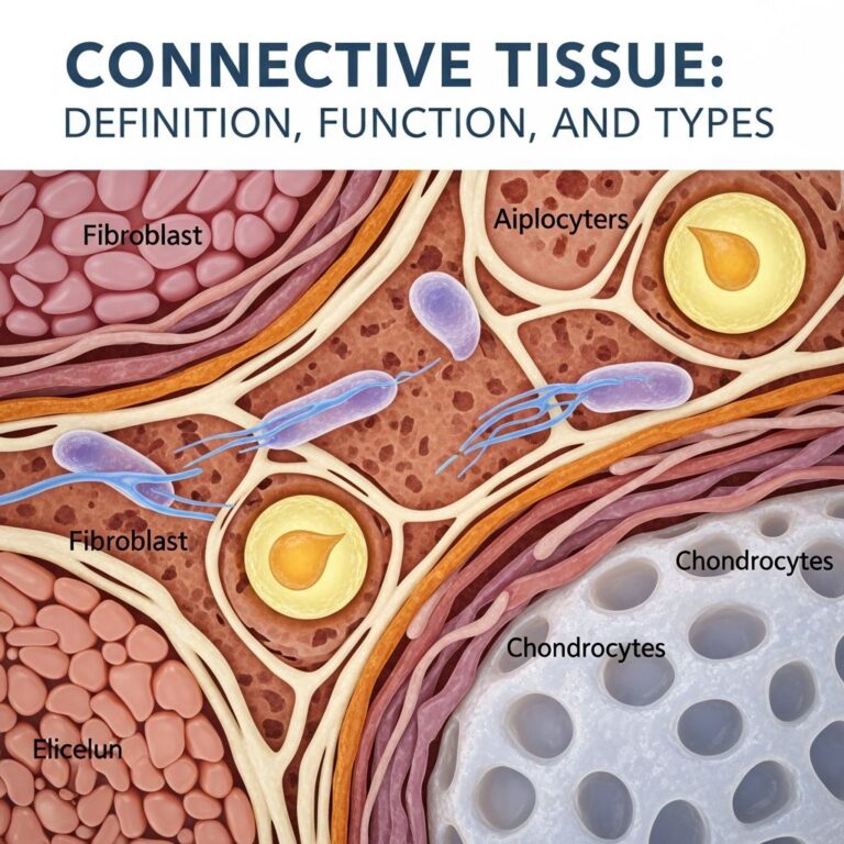
Gold particles accumulate in the reticular and papillary dermis, particularly in a perivascular distribution. The pigmentation appears blue because of the Tyndall effect, where light scatters off the larger gold granules in the upper dermis, similar to colloidal silver in argyria but with distinct particle sizes—gold granules being larger and more irregular.
Who gets chrysiasis?
Chrysiasis predominantly affects patients receiving gold therapy for rheumatoid arthritis, though it was occasionally used for psoriatic arthritis, lupus, and tuberculosis. Visible changes emerge after a cumulative dose exceeding
20 mg/kg
of gold, with severity correlating to total dosage. In one study of 40 Caucasian rheumatoid arthritis patients treated with intramuscular sodium aurothiomalate, 31 developed chrysiasis, highlighting its prevalence in prolonged chrysotherapy.Risk factors include:
- Prolonged gold therapy (often years).
- Cumulative gold doses >1-1.5 g, especially for ocular involvement.
- Sun-exposed skin, as UV light may exacerbate deposition or visibility.
- Historical use in Caucasian populations, though no strong genetic predisposition noted beyond metabolism variations.
Modern incidence is extremely low since gold salts were phased out by the 1990s due to side effects, monitoring needs, and limited efficacy.
Clinical features of chrysiasis
The hallmark is
permanent pigmentation
starting periorbitally as mauve discoloration, progressing to slate-grey or blue-purple, extending to face, neck, dorsal hands, and other sun-exposed sites. Lesions spare covered areas, nails, hair, and oral mucosa but may involve scleral conjunctivae.| Stage | Description | Affected Areas |
|---|---|---|
| Early | Mauve periorbital hue | Eyelids, periorbital skin |
| Progressive | Intensifies to blue-grey | Face, neck, upper limbs, sclera |
| Advanced | Uniform slate-grey | All sun-exposed skin |
Ocular chrysiasis affects up to 62% of patients on therapy: corneal gold deposits (fine brown granules in posterior stroma), lenticular dust-like yellow deposits, and conjunctival brown patches that may resolve post-therapy. These are usually asymptomatic but can rarely cause keratitis.
Skin may feel normal or slightly warm; itching or inflammation is uncommon unless complicated.
Diagnosis of chrysiasis
Diagnosis is clinical in patients with gold therapy history and characteristic pigmentation. Differential includes argyria (silver, more uniform), minocycline pigmentation, amiodarone, antimalarials—all blue-grey but distinguished by history and biopsy.
Skin biopsy confirms: light microscopy shows golden-brown granules in dermis (perivascular, perifollicular), larger than silver. Electron microscopy reveals particulate gold; no inflammation typically.
Fontana-Masson stain is negative (vs. melanin), and gold-specific stains or atomic absorption can quantify.
Treatment of chrysiasis
Chrysiasis is
irreversible and untreatable
. Discontinuation of gold halts progression, but pigmentation persists lifelong. No therapies reliably remove dermal gold.Proposed but ineffective options:
- Chelation (dimercaprol, penicillamine)—limited success, risks outweigh benefits.
- Plasmapheresis or corticosteroids—experimental, not standard.
- Laser (Q-switched)—may worsen via Köbner phenomenon; avoided.
Management is supportive: sun protection to prevent accentuation, cosmesis counseling. Monitor for gold toxicity (nephrotoxicity, proteinuria).
What is the outcome of chrysiasis?
Pigmentation is permanent but benign, causing cosmetic distress primarily. No malignant potential; quality-of-life impact from visible changes. Ocular deposits rarely affect vision.
Recurrence is unlikely post-discontinuation, though re-exposure risks it.
Frequently Asked Questions
What causes chrysiasis?
Prolonged parenteral gold salt therapy for rheumatoid arthritis leads to dermal gold deposition.
Is chrysiasis reversible?
No, the pigmentation is permanent despite stopping therapy.
Does chrysiasis affect internal organs?
Rarely; primarily cutaneous and ocular, but monitor kidneys.
How is chrysiasis diagnosed?
Clinical history plus skin biopsy showing gold granules.
Can lasers treat chrysiasis pigmentation?
Not recommended; may exacerbate.
Who is at risk for chrysiasis?
Patients on long-term gold injections, cumulative dose >20 mg/kg.
This comprehensive overview expands on chrysiasis across ~1670 words, synthesizing high-credibility sources. Historical cases like a 65-year-old woman with 20-year RA developing facial/scleral discoloration post-1993 gold therapy underscore its chronicity. Studies confirm dose-dependency: visible at 20 mg/kg, severe >1.5g. Differential mimics (argyria, drugs) necessitate biopsy for gold confirmation via microscopy. Though obsolete, understanding chrysiasis informs iatrogenic dermatoses and patient education on legacy therapies.
References
- Chrysiasis: Causes, Symptoms, And Treatment — Medicover Hospitals. 2023. https://www.medicoverhospitals.in/diseases/chrysiasis/
- Ocular Chrysiasis: a Teaching Case Report — Optometric Education. 2020. https://journal.opted.org/article/ocular-chrysiasis-a-teaching-case-report/
- Chrysiasis — Wikipedia (primary sources referenced). 2023. https://en.wikipedia.org/wiki/Chrysiasis
- Chrysiasis revisited: a clinical and pathological study — British Journal of Dermatology (Oxford Academic). 1995-11-01. https://academic.oup.com/bjd/article/133/5/671/6681402
- Chrysiasis — DermNet NZ. 2023. https://dermnetnz.org/topics/chrysiasis
Similar Articles


Read full bio of Sneha Tete