Coagulase-Negative Staphylococci: Clinical Features and Management
Comprehensive overview of coagulase-negative staphylococcal infections, diagnosis, and treatment strategies.

Introduction to Coagulase-Negative Staphylococci
Coagulase-negative staphylococci (CoNS) represent a diverse group of gram-positive, aerobic bacteria that are ubiquitous members of the normal human skin microbiota. The human skin serves as the first line of defense between the body and the external environment, and consequently hosts a rich microbial ecosystem comprising at least 47 species of coagulase-negative staphylococci. These organisms are distinguished from the closely related Staphylococcus aureus by their inability to produce coagulase, an enzyme that causes blood plasma to clot. Despite their abundance on intact skin, CoNS rarely cause disease in healthy individuals with intact skin barriers. However, the clinical significance of these organisms becomes apparent when breached skin surfaces occur or when medical implants provide surfaces for bacterial colonization and biofilm formation.
Microbiology and Characteristics
Coagulase-negative staphylococci are characterized as catalase-positive, gram-positive cocci that typically occur in clusters when viewed under microscopy. These organisms are predominantly found on the skin and mucous membranes, making them some of the most common bacteria colonizing human surfaces. The group comprises a heterogeneous collection of species, with over 30 identified species, and approximately 50% of these are known to colonize humans.
Major Pathogenic Species
Several CoNS species have particular clinical relevance:
- Staphylococcus epidermidis represents the most common CoNS species. Colonies typically appear small, white-beige in color, measuring approximately 1-2 mm in diameter. This species is particularly significant in healthcare settings as it frequently causes infections associated with indwelling medical devices.
- Staphylococcus saprophyticus is responsible for 10-15% of uncomplicated, community-acquired urinary tract infections, particularly in young women, making it clinically important for urinary tract pathology.
- Staphylococcus haemolyticus produces colonies that appear small and golden yellow, measuring about 1-2 mm in diameter. This species is notable for its frequent antibiotic resistance patterns.
- Staphylococcus lugdunensis forms colonies that are typically sticky, smooth, glossy, and yellow-orange in color, ranging from 2-4 mm in diameter. This species has emerged as a more virulent CoNS capable of causing serious infections.
Coagulase-Negative Staphylococcal Skin Conditions
While CoNS are normal skin inhabitants, certain strains and conditions can lead to dermatological manifestations. Understanding the relationship between CoNS and skin disorders is crucial for proper diagnosis and management.
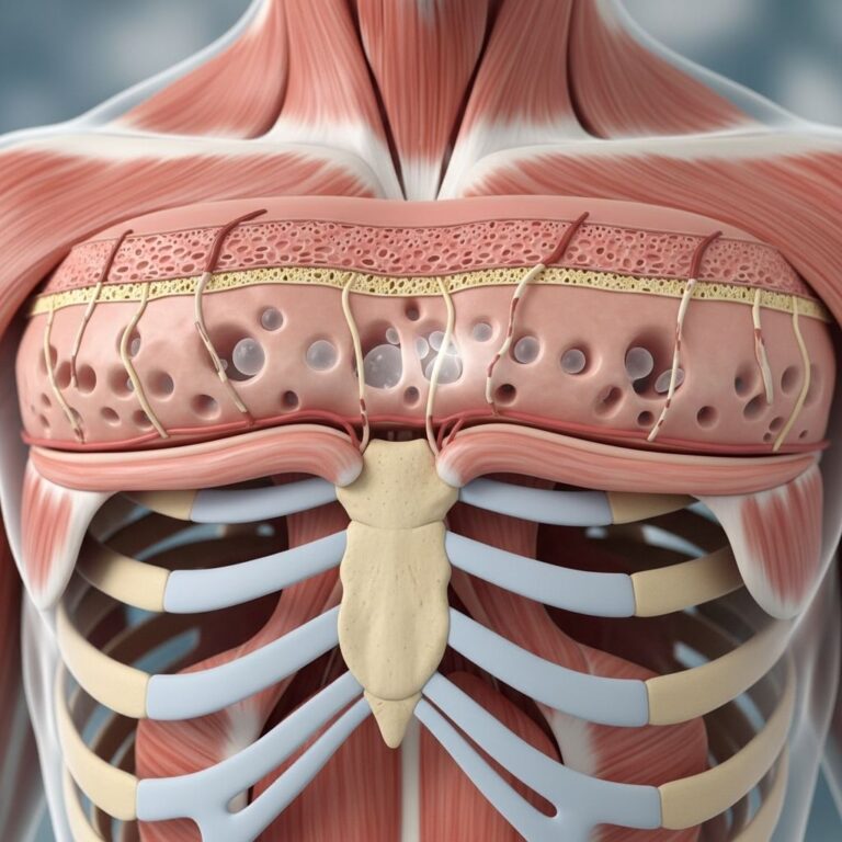
Miliaria and Sweat Duct Occlusion
S. epidermidis can induce miliaria, a disorder characterized by the retention of sweat within the eccrine glands. Skin biopsies from affected individuals have revealed that periodic acid-Schiff (PAS)-positive material tends to block the upper eccrine sweat ducts. This pathological process is specifically associated with extracellular polysaccharide (EPS)-producing strains of S. epidermidis. Notably, up to 62% of S. epidermidis strains isolated from the forehead and back produce EPS, indicating a substantial proportion of skin colonizers have this pathogenic capability.
Miliaria associated with CoNS is not produced by non-EPS producing strains of S. epidermidis or other coagulase-negative species such as S. haemolyticus and S. hominis. Occluded sweat ducts may also lead to additional complications including hyperhidrosis (excessive sweating) and anhidrosis (decreased sweating), which can occur in chronic dermatoses such as psoriasis, atopic dermatitis, and systemic sclerosis.
Role in Atopic Dermatitis
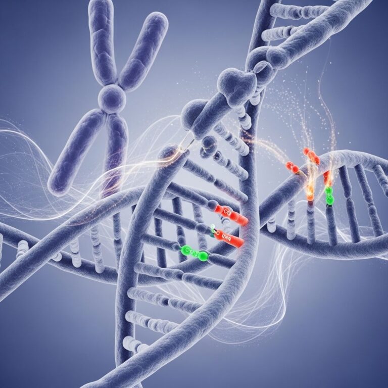
Coagulase-negative staphylococci are implicated in the "double-hit" phenomenon, a theoretical framework used to explain the pathogenesis of atopic dermatitis. This theory proposes that atopic dermatitis results from the combined effects of an abnormal stratum corneum (skin surface) and an unknown environmental trigger. The abnormal skin barrier is attributed to genetic factors, particularly mutations in the FLG gene, which encodes filaggrin, a crucial protein for skin barrier function.
Research has identified that the specific environmental trigger in atopic dermatitis may be subclinical miliaria caused by CoNS. PAS-positive material has been discovered in the eccrine ducts of patients with atopic dermatitis, and this material may arise from both S. aureus and EPS-producing strains of S. epidermidis. Rather than causing typical miliaria lesions, in patients with an FLG defect, occlusion of the eccrine ducts may trigger a flare of atopic dermatitis by activating the innate immune system.
Interestingly, skin colonization patterns differ between healthy individuals and those with atopic dermatitis. Skin samples from patients with atopic dermatitis are colonized with a greater proportion of S. aureus than healthy controls, which are typically colonized with S. epidermidis. Evidence suggests that skin colonization by S. epidermidis may confer protection against atopic dermatitis, particularly in patients with the FLG gene defect. This protective role reflects the competitive antagonism between different staphylococcal species on the skin surface.
Risk Factors and Pathogenesis of Infection
Despite their abundance on the skin, coagulase-negative staphylococci rarely cause disease in individuals with intact skin barriers. The primary risk factor for coagulase-negative staphylococcal infection is the presence of a medical implant, such as central venous catheters, prosthetic joints, artificial heart valves, pacemakers, or other indwelling devices. On these foreign surfaces, the organisms can colonize, proliferate, and potentially gain access to the systemic circulation.
Mechanism of Infection
CoNS typically gain entry through breached skin surfaces, commonly during medical or nursing procedures. The key pathogenic mechanism is the remarkable ability of these bacteria to form biofilms on the surfaces of implanted medical equipment. Biofilms are organized communities of bacteria encased in an extracellular matrix that provides protection from the immune system and antibiotics. Within biofilms, bacteria replicate and can disseminate into the systemic circulation, causing bacteremia and potentially life-threatening sepsis.
The process of infection involves several key steps: initial adhesion to the medical device surface, followed by biofilm maturation, bacterial replication within the protected microenvironment, and eventual dissemination to other body sites. This biofilm-based pathogenesis makes CoNS infections particularly challenging to treat, as the bacteria within biofilms exhibit reduced susceptibility to antimicrobial agents.
Demographics and Clinical Presentations
Coagulase-negative staphylococcal infections predominantly occur in healthcare settings and affect specific patient populations at elevated risk.
Vulnerable Patient Populations
Several groups are particularly susceptible to CoNS infections:
- Hospitalized patients with central venous catheters or other indwelling medical devices represent the largest affected population in healthcare settings.
- Immunocompromised and immunosuppressed patients, including those with human immunodeficiency virus (HIV), organ transplant recipients, and patients receiving chemotherapy, face significantly elevated risk.
- Critically ill patients in intensive care units who often have multiple indwelling devices and breached skin barriers.
- Premature infants are particularly vulnerable to CoNS-induced infections, including pneumonia, omphalitis, and skin abscesses. Preterm neonates are especially susceptible to developing sepsis from CoNS bacteremia.
- Older adults with significant comorbidities demonstrate increased risk compared with the general population.
Clinical Manifestations by Site of Infection
| Site of Infection | Clinical Presentation | Associated Risk Factors |
|---|---|---|
| Central venous catheter | Fever, chills, bacteremia, sepsis | Prolonged catheterization, immunosuppression |
| Prosthetic joints | Joint pain, swelling, erythema, drainage | Recent orthopedic surgery, immunodeficiency |
| Peritoneal dialysis catheter | Fever, nausea, vomiting, abdominal pain and tenderness | Long-term dialysis, procedural complications |
| Endophthalmitis | Redness, pain, impaired vision | Intravitreal injection, eye trauma |
| Urinary tract infection | Pyuria, often asymptomatic | Urinary catheterization, particularly S. saprophyticus |
| Genitourinary prostheses | Penile erythema, swelling, pain, induration | Prosthetic implantation |
| Breast implants and mastitis | Localized pain, erythema, swelling, tenderness, drainage | Implant presence, procedural complications |
| Cardiac devices | Fever, signs of sepsis, device malfunction | Pacemaker or implantable cardioverter-defibrillator placement |
Diagnosis of Coagulase-Negative Staphylococcal Infections
Diagnosing CoNS infections presents clinical challenges because these organisms are normal skin inhabitants and frequently contaminate cultures. Proper diagnostic interpretation requires clinical correlation and careful specimen collection techniques.
Culture and Identification
CoNS grow readily on standard bacterial culture media including blood agar and chocolate agar. Colonial morphology varies by species and can aid in preliminary identification. Once isolated, definitive identification relies on biochemical testing to confirm the absence of coagulase production, distinguishing CoNS from the coagulase-positive S. aureus.
Distinguishing Infection from Contamination
Multiple positive blood cultures with CoNS from different collection sites, growth from normally sterile body fluids, or isolation from prosthetic device samples more reliably indicate true infection rather than contamination. Clinical context, including fever, signs of sepsis, and device-related symptoms, must be considered when interpreting culture results.
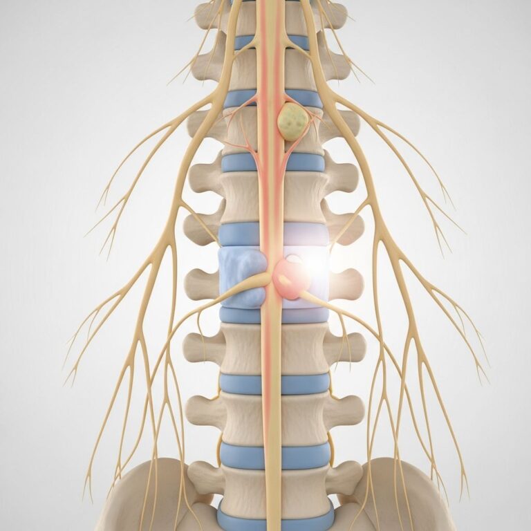
Central Nervous System Shunt Infections
Central nervous system (CNS) shunt infections caused by CoNS represent a particularly serious form of device-related infection. These infections can lead to significant morbidity and mortality if not promptly diagnosed and treated. S. epidermidis is the most common cause of CNS shunt infections, reflecting its prevalence on skin surfaces where shunt catheters breach the barrier.
Clinical manifestations of CNS shunt infections include fever, headache, meningeal signs, altered mental status, and signs of increased intracranial pressure. Cerebrospinal fluid (CSF) analysis typically shows elevated protein levels, normal or low glucose, and pleocytosis. Treatment of CNS shunt infections requires both systemic antibiotics with CNS penetration and, in most cases, removal or external drainage of the infected shunt.
Antibiotic Susceptibility and Treatment
Antibiotic resistance represents a serious and common challenge in treating CoNS infections. Understanding the resistance patterns of these organisms is essential for selecting appropriate therapy.
Resistance Patterns
Approximately 90% of CoNS infections are resistant to penicillin, and greater than 80% are resistant to methicillin (oxacillin and nafcillin). This widespread resistance is mediated by the mecA gene, which encodes a low-affinity penicillin-binding protein. CoNS are frequently resistant to multiple antibiotic classes including ciprofloxacin, gentamicin, erythromycin, and clindamycin, requiring careful antimicrobial stewardship.
Importantly, resistance patterns can be heterotypic, meaning that individual bacterial isolates from the same patient may demonstrate different susceptibility patterns. Therefore, obtaining multiple isolates and performing comprehensive susceptibility testing is recommended to determine whether organisms are truly susceptible to beta-lactam antibiotics.
Recommended Antibiotic Therapy
Vancomycin is considered the drug of choice for serious CoNS infections, particularly those caused by methicillin-resistant strains. For organisms confirmed to be susceptible to methicillin, vancomycin can be replaced by methicillin-resistant penicillin (nafcillin), first-generation cephalosporins (such as cefazolin), or second-generation cephalosporins (such as cefoxitin).
Newer antibiotics with proven activity against CoNS include daptomycin, linezolid, clindamycin, telavancin, tedizolid, and dalbavancin. These agents may be considered for patients with documented susceptibility or in cases where standard therapy is contraindicated. For deep-seated infections, gentamicin or rifampicin can be added to enhance bacterial killing and penetration into biofilm.
Duration of Therapy
The mainstay of treatment for CoNS infections involves appropriate systemic antibiotic therapy combined with removal of the culprit implant whenever possible. Device removal is critical because bacteria within biofilms on prosthetic materials exhibit significantly reduced antibiotic susceptibility, and prolonged antimicrobial therapy alone is unlikely to achieve cure without removing the infected foreign body.
Decolonization Strategies
Due to the correlation between mucosa colonization and subsequent bacteremia, preventive treatment can be administered to decrease mucosal colonization with CoNS. One suggested approach includes topical mupirocin for nasal decolonization and an oral glycopeptide, such as ramoplanin, for intestinal decolonization. These strategies may be particularly valuable in preventing recurrent infections in high-risk patients with recurrent CoNS bacteremia.
Clinical Outcomes and Prognosis
Coagulase-negative staphylococcal bacteremia is a serious medical condition associated with significant morbidity and mortality, particularly in hospitalized and immunocompromised patients. While superficial CoNS infections of intact skin are rare and generally benign, systemic infections require aggressive management with appropriate antibiotics and device removal.
Outcomes depend on multiple factors including the severity of underlying illness, adequacy of immune function, timely diagnosis, appropriate antibiotic selection, and removal of infected prosthetic material. Early recognition and treatment of CoNS infections significantly improve patient outcomes and reduce complications such as septic shock, disseminated infection, and device failure.
Frequently Asked Questions
Q: Are coagulase-negative staphylococci always pathogenic?
A: No. CoNS are normal skin commensals and rarely cause disease in individuals with intact skin. They typically only cause infection when medical implants are present or when the skin barrier is significantly compromised, allowing bacterial entry into deeper tissues or the bloodstream.
Q: Why are medical devices particularly susceptible to CoNS infection?
A: CoNS possess a remarkable ability to produce biofilms on prosthetic material surfaces. These biofilms protect bacteria from immune system attack and significantly reduce antibiotic penetration, making infections difficult to treat and necessitating device removal for cure.
Q: Can CoNS infections be treated with penicillin or methicillin?
A: Most CoNS infections (approximately 90%) are resistant to penicillin, and over 80% are resistant to methicillin. Vancomycin is typically the drug of choice for resistant infections, though susceptibility testing is essential for guiding therapy.
Q: Why is device removal important in treating CoNS infections?
A: Bacteria within biofilms on prosthetic devices exhibit markedly reduced antibiotic susceptibility. Removal of the infected device is crucial because antibiotic therapy alone, without device removal, is unlikely to achieve complete cure.
Q: Are certain CoNS species more pathogenic than others?
A: Yes. Staphylococcus lugdunensis is considered more virulent and capable of causing more serious infections compared to other CoNS species. Staphylococcus saprophyticus is particularly important for causing urinary tract infections, especially in young women.
References
- Coagulase-Negative Staphylococci — DermNet New Zealand. Updated 2024. https://dermnetnz.org/topics/coagulase-negative-staphylococci
- Coagulase-Negative Staphylococcus — Infectious Disease Advisor, Springer Healthcare. 2024. https://www.infectiousdiseaseadvisor.com/ddi/coagulase-negative-staphylococcus/
- Staphylococci, Coagulase Negative — Johns Hopkins ABX Guide, Johns Hopkins University. 2024. https://www.hopkinsguides.com/hopkins/view/Johns_Hopkins_ABX_Guide/540517/all/Staphylococci__coagulase_negative
- Coagulase-Negative Staphylococcal Infections — Red Book: 2024-2027 Report of the Committee on Infectious Diseases, American Academy of Pediatrics. https://publications.aap.org/redbook/book/755/chapter/14081883/Coagulase-Negative-Staphylococcal-Infections
Similar Articles


Read full bio of medha deb