Computed Tomography Angiography (CTA): Complete Guide
Comprehensive guide to CTA imaging: procedure, uses, benefits, and what to expect.
: Complete Guide")
What is Computed Tomography Angiography (CTA)?
Computed tomography angiography (CTA), also known as CT angiography, is a non-invasive diagnostic imaging procedure that combines computed tomography technology with contrast material injection to create detailed, high-resolution images of blood vessels throughout the body. This advanced imaging technique uses X-ray technology and sophisticated computer processing to visualize arteries and veins with exceptional clarity and precision. CTA allows healthcare providers to assess the structure and function of blood vessels, identify abnormalities, and plan appropriate treatment strategies for various vascular conditions.
Unlike traditional catheter angiography, which is an invasive procedure requiring a catheter to be threaded through blood vessels, CTA is minimally invasive and can typically be performed in an outpatient radiology department or imaging center. The procedure has revolutionized vascular imaging by providing submillimeter spatial resolution and high temporal resolution, enabling physicians to detect even subtle vascular abnormalities.
How CTA Works
The CTA procedure utilizes multiple X-ray beam sources and detectors that spin around the body at high speed. As these X-ray beams pass through the body, specialized detectors capture the radiation that exits, creating cross-sectional images. A fast computer then processes this large volume of data to produce two-dimensional and three-dimensional images of the blood vessels and surrounding tissues.
A key component of CTA imaging involves the use of iodinated contrast material (ICM), which is injected intravenously to enhance the visibility of blood vessels. The contrast material flows through the bloodstream, making arteries and veins appear bright on the CT images. Modern CT scanners can provide high-quality three-dimensional datasets within a single breath-hold during the first pass of the contrast material, allowing for excellent image quality with minimal motion artifacts.
The timing of scanning in relation to contrast injection is crucial for CTA success. A test bolus technique involves injecting a small amount of contrast to measure how quickly it travels through the blood vessels. Once this transit time is determined, the full contrast bolus is injected and scanning begins at the optimal timing, ensuring that the vessels of interest are captured at peak enhancement.
CTA Imaging Protocols and Techniques
A typical CTA protocol includes multiple phases of imaging to ensure comprehensive evaluation of vascular pathology. These phases include:
Non-Contrast Phase: This initial imaging phase is particularly valuable in specific clinical scenarios. For patients with suspected acute aortic syndrome, non-contrast images help evaluate the presence of aortic intramural hematoma. In patients with suspected bleeding, non-contrast images can identify areas of hemorrhage. Additionally, non-contrast imaging is useful for differentiating calcifications and surgical graft material from contrast enhancement or potential graft leaks.
Arterial Phase: During the arterial phase, contrast material fills the arteries, providing optimal visualization of arterial structures and disease. This phase is essential for detecting arterial stenosis, blockages, and aneurysms.
Delayed Phase: The delayed phase imaging occurs after contrast has passed through the arterial system and entered the venous circulation. This phase helps assess venous structures and can reveal certain pathologies that may not be apparent on earlier phases.
Modern CTA requires specific technical requirements for optimal performance. A 64-channel CT scanner is considered the minimum requirement for advanced CTA applications such as coronary CTA, while a 16-channel CT system is typically adequate for other vascular applications such as abdominal aorta or visceral aneurysm assessment.
Clinical Applications of CTA
CTA has become an indispensable diagnostic tool for evaluating a wide range of vascular conditions across multiple organ systems.
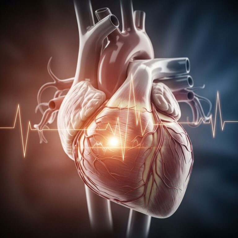
Coronary Artery Imaging
Coronary CT angiography (CCTA) represents one of the most significant applications of this technology. The procedure involves intravenous injection of contrast material followed by high-speed CT scanning of the heart. With modern CT technology, patients can typically be scanned without requiring heart-slowing medications, simply by holding their breath during the scan. CCTA is used to assess irregularities in heart and vessel structure, determine the location of stents and verify their patency, and screen for atherosclerotic disease. Many patients can now undergo CCTA instead of conventional catheter angiography, though the latter has not been completely replaced. CCTA is particularly valuable for early detection of arterial narrowing, enabling timely corrective therapy. The procedure is safer, faster, and more cost-effective than traditional catheter angiography.
Aortic and Great Vessel Imaging
CTA is the test of choice for identifying aneurysms in the aorta and other major blood vessels. These areas of weakened vessel wall that bulge outward can be life-threatening if rupture occurs. CTA provides superior ability to detect calcium deposits within vessel walls and allows better estimation of vessel dilation compared to standard angiography. Additionally, CTA excels at detecting blood clots within aneurysms. CTA is also used to identify arterial dissection, where the layers of the artery wall peel away from each other, potentially causing pain and life-threatening complications. The procedure quickly and non-invasively identifies dissections, demonstrates disease extent, and reveals any associated leakage.
Peripheral Vascular Disease
CTA is essential for localizing and confirming hemodynamically significant stenosis in patients with atherosclerosis who require surgical or endovascular procedures. The imaging provides a detailed roadmap of vascular lesions, guiding physicians in selecting the appropriate procedure and access route. Fixed luminal stenosis, the most common outcome of atherosclerotic disease, can limit blood flow and result in symptoms such as claudication (leg pain with walking), angina (chest pain), renovascular hypertension, and abdominal angina. CTA can detect plaque disease that has narrowed arteries to the legs and helps prepare patients for angioplasty, stent placement, or surgery.
Renal Artery Assessment
CTA can detect disease in the renal arteries and visualize blood flow to help prepare patients for kidney transplantation or renal artery stent placement. This application is particularly important for evaluating renovascular hypertension and assessing kidney perfusion prior to transplantation.
Pulmonary Embolism Detection
CTA can examine pulmonary arteries to detect pulmonary embolism (blood clots such as those traveling from leg veins) and identify pulmonary arteriovenous malformations. This represents a critical application in emergency medicine for rapidly diagnosing this life-threatening condition.
Additional Applications
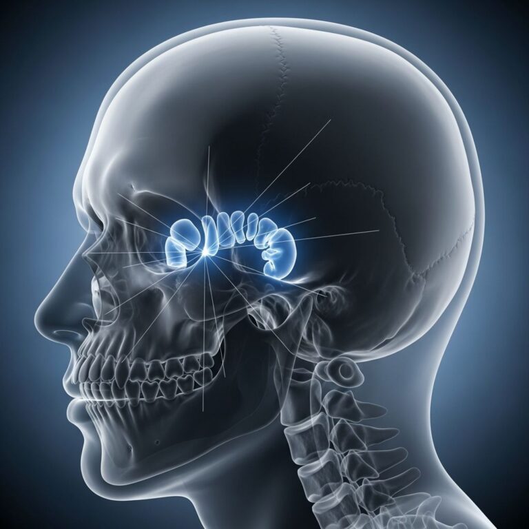
CTA has numerous other clinical applications, including identifying arteriovenous malformations in the brain or elsewhere, detecting injuries to arteries following trauma, evaluating arteries feeding tumors prior to surgery or other therapeutic procedures, assessing congenital blood vessel abnormalities in children, and guiding interventional radiologists and surgeons during repairs to diseased blood vessels.
Advantages and Benefits of CTA
CTA offers multiple advantages over alternative imaging modalities and diagnostic approaches. The procedure provides superior anatomical detail compared to magnetic resonance imaging (MRI) or ultrasound. CTA is minimally invasive with significantly reduced risk compared to conventional catheter angiography. The procedure is rapid, allowing most examinations to be completed in one to two minutes, though multiple scans may be required in some cases. CTA is cost-effective and can typically be performed in an outpatient setting without hospital admission. The three-dimensional imaging capabilities enable comprehensive assessment of complex vascular anatomy and pathology. Additionally, CTA provides accurate bolus timing techniques essential for high image quality.
Radiation Dose Reduction Techniques
Modern CTA incorporates several advanced techniques to minimize radiation exposure while maintaining excellent image quality. These innovative approaches include:
Low kV Technique: Reducing the kilovoltage (kV) settings decreases radiation dose while maintaining diagnostic image quality.
Iterative Reconstructions: Advanced reconstruction algorithms reduce image noise and allow for lower radiation dose protocols.
Hybrid Photon Imaging (HPH): This emerging technology improves image quality while reducing radiation exposure.
Dual Energy CT (DECT): Dual energy imaging allows for better tissue characterization and can reduce both radiation dose and contrast requirements in appropriate clinical scenarios.
What to Expect During Your CTA Procedure
Understanding the CTA procedure can help reduce patient anxiety and improve cooperation during imaging. During the examination, you will lie on a table that moves through the CT scanner opening as X-ray beams examine different areas of your body. A healthcare technologist will place an intravenous (IV) catheter, typically in your arm, before the procedure begins. An automatic injection pump connected to the IV will inject contrast material at a specified rate. In certain cases, particularly with children or patients with fragile or small veins, contrast material may be hand-injected using a syringe.
During scanning, the table is positioned at the imaging start point and then moves through the machine opening as the actual CT scanning is performed. For coronary CTA, you may be asked to hold your breath during the scan to minimize motion artifacts. The entire procedure typically takes only a few minutes from start to finish.
Safety Considerations and Contrast Material
CTA uses iodinated contrast material to enhance blood vessel visualization. While contrast material is generally safe, some patients may experience minor side effects such as a warm sensation or metallic taste. Serious allergic reactions are rare but possible. Patients with certain medical conditions, such as severe kidney disease, diabetes, or iodine allergies, should inform their healthcare provider before undergoing CTA so appropriate precautions can be taken.
The procedure exposes patients to ionizing radiation, though the dose is typically minimal and the benefits generally outweigh the risks for appropriate clinical indications. Modern CTA protocols incorporate radiation dose reduction techniques to minimize exposure.
CTA in Surgical and Interventional Planning
CTA serves important roles beyond diagnosis, including pre-operative and pre-interventional planning. The detailed vascular roadmap provided by CTA helps surgeons and interventional radiologists plan complex vascular procedures. CTA is often utilized prior to surgical procedures requiring vascular reconstruction or graft placement, such as those in organ transplantation. CTA may serve as a scouting procedure to define vascular anatomy in high-risk surgeries such as coronary artery bypass grafting (CABG) or redo sternotomy.
Post-Processing and Image Analysis
After completion of scanning, advanced post-processing techniques are employed to optimize image quality and visualization. Three-dimensional image reconstruction can be created from the raw CT data, allowing physicians to view vascular structures from multiple angles and perspectives. These 3D reconstructions provide enhanced understanding of complex vascular anatomy and pathology, facilitating better clinical decision-making and patient communication.
Frequently Asked Questions About CTA
Q: Is CTA painful?
A: CTA is painless. You may feel slight discomfort from IV placement, and some patients report a warm sensation or metallic taste when contrast material is injected. The scanning itself is completely painless.
Q: How long does a CTA procedure take?
A: A single CTA scan typically takes one to two minutes, though the entire appointment including preparation and post-scan review may take 30-60 minutes total.
Q: Can I eat or drink before my CTA?
A: Instructions vary depending on the specific type of CTA being performed. Your healthcare provider will give you specific pre-procedure instructions. In general, you may need to fast for a few hours before the procedure.
Q: What are the risks of CTA?
A: CTA is very safe overall. Potential risks include allergic reactions to contrast material (rare), kidney issues in susceptible patients, and radiation exposure. Serious complications are uncommon when the procedure is performed by experienced radiologists.
Q: Is CTA better than MRI for vascular imaging?
A: CTA provides superior anatomical detail compared to MRI and is faster, making it ideal for emergency situations. However, MRI may be preferred in certain cases. Your physician will determine the best imaging modality for your specific condition.
Q: Can CTA replace cardiac catheterization?
A: Many patients can undergo CCTA instead of conventional catheter angiography, as it is safer, faster, and more cost-effective. However, catheterization has not been completely replaced, as some clinical situations still require its use.
Q: What should I do after my CTA?
A: After CTA, you can typically resume normal activities immediately. Drink plenty of water to help flush the contrast material from your system. Your radiologist will provide a report to your physician within 24 hours.
References
- Computed Tomography Angiography (CTA) | Clinical Keywords — Yale Medicine. https://www.yalemedicine.org/clinical-keywords/computed-tomography-angiography
- Vascular computed tomography angiography technique and clinical applications — National Center for Biotechnology Information (NCBI). 2019. https://pmc.ncbi.nlm.nih.gov/articles/PMC6732113/
- Computed tomography angiography — Wikimedia Foundation. https://en.wikipedia.org/wiki/Computed_tomography_angiography
- CT Angiogram (CTA) — Radiology — University of California, San Francisco Health. https://radiology.ucsf.edu/patient-care/services/ct-angiography
- CT Angiography (CTA) — Radiology Info. https://www.radiologyinfo.org/en/info/angioct
Similar Articles


Read full bio of medha deb