Congenital Melanocytic Naevi: Guide To Diagnosis, Risks & Care
Comprehensive guide to congenital melanocytic naevi: from diagnosis and risks to management strategies for patients and families.

Congenital melanocytic naevi (CMN), also known as congenital melanocytic nevi, are pigmented skin lesions present at birth caused by proliferation of melanocytes, the pigment-producing cells. These naevi range from small tan patches to giant, darkly pigmented areas covering large body surfaces and carry risks of malignant transformation and neurological complications.
What are congenital melanocytic naevi?
Congenital melanocytic naevi are benign proliferations of melanocytes appearing as brown, black, or bluish patches at birth or within the first few weeks of life. They result from a postzygotic somatic mutation, typically in genes like NRAS, leading to localized melanocyte hyperplasia during fetal development. Prevalence is approximately 1% of newborns, with smaller naevi more common and giant forms rarer (1 in 20,000 births).
CMN are classified by projected adult size: small (<1.5 cm), medium (1.5–20 cm), and
giant
(>20 cm), also termed garment, bathing trunk, or hairy naevi when extensive. Giant CMN often involve the trunk and may have satellite lesions—smaller CMN elsewhere on the body.Who gets congenital melanocytic naevi?
CMN occur sporadically without familial inheritance, affecting all ethnicities equally. Risk factors include posterior axial location (back) for giant CMN and multiple satellite lesions, correlating with higher complication risks. Males and females are equally affected.
What causes congenital melanocytic naevi?
The primary cause is somatic mosaicism from mutations in melanocyte lineage genes, most commonly NRAS (70–80% of cases), leading to uncontrolled melanocyte proliferation in the skin and sometimes leptomeninges. These mutations occur post-conception, explaining lack of heritability. Environmental factors are not implicated.
What are the clinical features of congenital melanocytic naevi?
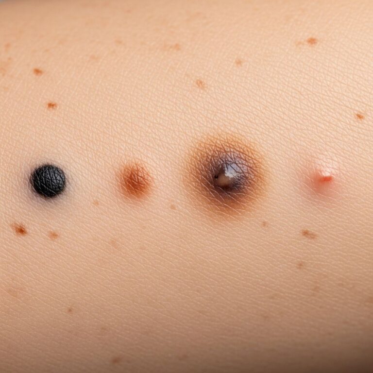
At birth, CMN appear as well-defined, flat, light to dark brown patches, often velvet-like or pebbly. Common sites include trunk (60–70%), limbs, and head/neck (15%).
- Colour: Tan-brown to black; may darken or variegate with age.
- Surface: Smooth, wrinkled, verrucous (warty), or nodular; hypertrichosis (hairiness) develops in 40–80%.
- Size: Varies; giant CMN cover >20 cm adult size, often with satellites (up to hundreds).
- Distribution: Single or multiple; posterior midline increases neurocutaneous risk.
Over time, naevi grow proportionally, develop nodules (neurotization), and may itch or ulcerate. Microscopically, melanocytes extend deeply into dermis, around adnexa, vessels, and fat—deeper than acquired naevi.
Diagnosis
Diagnosis is clinical based on history (present at birth) and appearance. Dermoscopy shows atypical pigment networks, globules, and hairs. Biopsy is rarely needed unless malignancy suspected, showing expanded melanocyte nests deeper in dermis. MRI brain/spine is recommended for giant CMN (>20 cm), posterior location, or >20 satellites to screen for neurocutaneous melanosis.
Classification of congenital melanocytic naevi
| Type | Projected Adult Size | Prevalence | Risk Profile |
|---|---|---|---|
| Small | <1.5 cm | ~1% | Low melanoma risk (<1%) |
| Medium | 1.5–20 cm | 1:100 | Moderate risk |
| Giant | >20 cm | 1:20,000 | High risk (5–10% melanoma) |
Classification guides risk stratification and management.
Complications
CMN complications span cosmetic, functional, and malignant domains.
- Cosmetic/Psychosocial: Visible naevi cause distress, bullying, anxiety.
- Local: Itching, ulceration, infection, especially in flexures.
- Musculoskeletal: Associated scoliosis, limb asymmetry in giant trunk naevi.
Neurocutaneous melanosis (NCM)
NCM involves melanocyte proliferation in leptomeninges, affecting 2–45% with giant CMN or numerous satellites. Symptomatic NCM (15–30%) presents with seizures, hydrocephalus, developmental delay. Asymptomatic cases found on MRI. Risk highest in posterior axial giant CMN.
Melanoma risk
Lifetime melanoma risk: small CMN <1%, giant 3–9% (17-fold general population increase). Melanomas may be cutaneous (within naevus), extracutaneous (CNS, viscera), or both. Risk peaks in childhood for NCM-related, adulthood for cutaneous. Nodular growth, ulceration, color change signal danger.
Management
Multidisciplinary approach: dermatology, plastic surgery, neurology, oncology, psychology. Goals: surveil for malignancy, treat symptoms, optimize cosmesis.
- Surveillance: Annual exams; photos, dermoscopy. MRI at diagnosis for high-risk, then 3–6 monthly if NCM suspected.
- Surgery: Complete excision ideal for small/medium; staged for giant using tissue expanders, grafts, flaps. Curettage in infancy for small lesions. Not always feasible due to size/recurrence.
- Laser: Q-switched for pigment; CO2 for texture—limited efficacy, recurrence common.
- Symptom relief: Emollients, steroids for itch/ulceration.
- Psychosocial: Counseling, camouflage makeup, support groups.
Prophylactic excision reduces local risk but not NCM melanoma. Shared decision-making weighs risks/benefits.
Prognosis
Excellent for small CMN. Giant CMN prognosis depends on NCM/melanoma. Symptomatic NCM mortality >50%; early detection improves outcomes. Surgical scarring may exceed naevus burden. Long-term surveillance lifelong.
Frequently Asked Questions
Are congenital melanocytic naevi cancerous?
Most are benign, but giant CMN carry 3–9% lifetime melanoma risk, higher than general population.
Should CMN be removed?
Small visible/symptomatic: yes. Giant: consider staged excision if feasible; surveillance alternative.
Does CMN mean my child has NCM?
Only 2–45% giant CMN have radiological NCM; 15–30% symptomatic. MRI screens high-risk cases.
Can CMN be prevented?
No, sporadic mutations; not inherited.
What changes warrant urgent review?
Nodular growth, bleeding, ulceration, color/texture change, pain.
Guidelines
- Annual dermatology review for all CMN.
- MRI brain/spine for giant CMN, >20 satellites, posterior location.
- Biopsy suspicious areas.
- Multidisciplinary team for giant CMN.
References
- Congenital Melanocytic Naevus: Features & Management — The Plastics Fella. 2023. https://www.theplasticsfella.com/congenital-melanocytic-naevus/
- Congenital Melanocytic Nevi (CMN) — Nationwide Children’s Hospital. 2024-01-15. https://www.nationwidechildrens.org/conditions/congenital-melanocytic-nevi
- Giant congenital nevus — MedlinePlus (U.S. National Library of Medicine). 2025. https://medlineplus.gov/ency/article/001453.htm
- Congenital Melanocytic Nevi — StatPearls (NCBI Bookshelf, NIH). 2024-07-20. https://www.ncbi.nlm.nih.gov/books/NBK563168/
- What are congenital melanocytic nevi? — Society for Pediatric Dermatology. 2023. https://pedsderm.net/site/assets/files/1028/spd_cmn_bw.pdf
- Congenital Nevi (Moles) in Children — Boston Children’s Hospital. 2024. https://www.childrenshospital.org/conditions-treatments/congenital-nevi-moles-children
Similar Articles


Read full bio of medha deb