Corynebacterial Skin Infections: Causes, Diagnosis & Treatment
Understanding corynebacterial infections: From pitted keratolysis to erythrasma and trichomycosis.

Introduction to Corynebacteria
Corynebacteria are small gram-positive cocci that are generally considered commensal organisms residing on normal human skin. However, under specific circumstances involving environmental factors such as heat, moisture, and occlusion, these bacteria can become pathogenic and cause clinically significant skin infections. Understanding the conditions that transform these normally benign organisms into disease-causing pathogens is essential for accurate diagnosis and effective treatment. This comprehensive guide explores the major corynebacterial skin infections, their clinical presentations, diagnostic approaches, and evidence-based management strategies.
Pitted Keratolysis: Clinical Features and Management
Overview and Pathophysiology
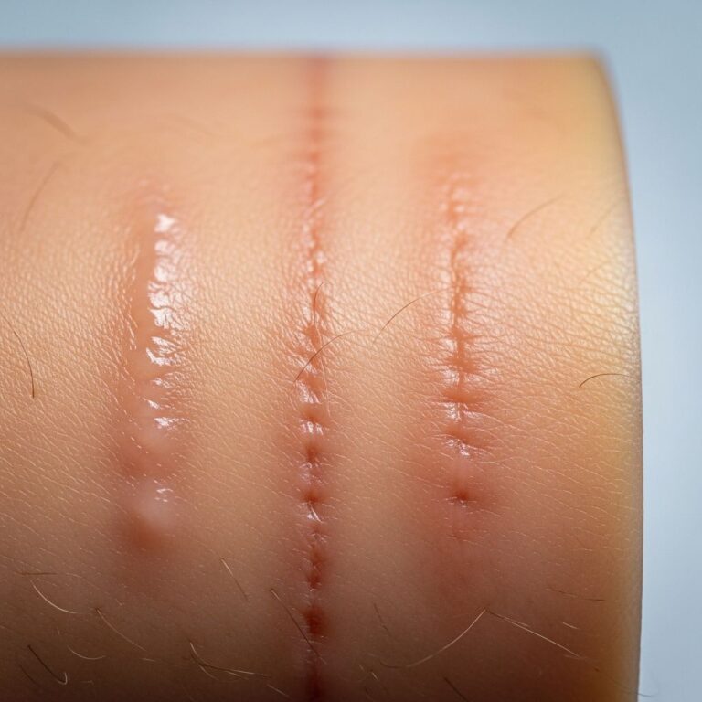
Pitted keratolysis is a superficial bacterial infection that primarily affects the soles of the feet, characterized by distinctive small, punctate erosions or pits in the stratum corneum. This condition is caused by corynebacteria and possibly Dermatophilus congolensis, with the infection being directly associated with specific environmental and host factors. The disease is strongly linked to hyperhidrosis (excessive sweating) and the prolonged use of occlusive footwear, which creates the warm, moist microenvironment necessary for bacterial proliferation.
Clinical Presentation
The most characteristic feature of pitted keratolysis is the appearance of the feet. Either the forefoot or the heel, or both areas simultaneously, become affected with white patches containing clusters of punched-out pits that create a distinctive appearance. These pits become more obvious and visually prominent when the skin is wet, making the diagnosis particularly apparent after bathing or swimming. The infected areas typically have a distinctive appearance that can be readily distinguished from other common foot conditions.
One of the most bothersome symptoms associated with pitted keratolysis is the production of a characteristic foul odor emanating from the feet. This malodorous quality often becomes the chief complaint driving patients to seek medical attention and can significantly impact quality of life and social interactions.
Treatment Approach
Pitted keratolysis responds well to topical antibacterial agents. Treatment options include:
- Topical antibiotics: Fusidic acid cream is particularly effective and represents a first-line topical treatment option.
- Oral antibiotics: Erythromycin administered orally can be used for more extensive or resistant cases, demonstrating rapid response in most patients.
However, without addressing the underlying predisposing factors, pitted keratolysis will quickly recur after successful treatment. Patient counseling and preventive measures are therefore essential components of long-term management. Practical advice should include:
- Maintaining feet in a dry state through frequent drying and moisture management
- Avoiding prolonged use of occlusive or airtight footwear
- Choosing breathable footwear materials that allow moisture evaporation
- Reducing sweating through antiperspirant use when appropriate
- Regular foot hygiene practices
Erythrasma: A Chronic Superficial Bacterial Infection
Etiology and Epidemiology
Erythrasma is a chronic, superficial bacterial infection caused by Corynebacterium minutissimum, a gram-positive, non-spore-forming, aerobic or facultative bacillus. The bacterium exists as part of the normal skin flora in many individuals but becomes pathogenic under conditions of heat, moisture, and occlusion. This infection occurs most commonly in specific anatomical locations, particularly in intertriginous areas where skin surfaces remain in close contact and moisture accumulates. The condition is notably prevalent in diabetic patients, where metabolic alterations and increased skin moisture create an ideal environment for bacterial proliferation.
Clinical Presentation
Erythrasma typically presents as a slowly enlarging area of pink or brown dry skin, most commonly affecting the axillae (armpits), groins, under the breasts, and between the toes. The infection is characteristically asymptomatic in many cases, though some patients may experience itching or mild discomfort. The lesions are well-defined with reddish-brown scaly patches that may gradually expand over time if left untreated.
Diagnostic Methods
Accurate diagnosis of erythrasma can be challenging, as its clinical appearance overlaps with other common dermatological conditions including tinea versicolor and candidiasis. Several diagnostic approaches can confirm the diagnosis:
- Wood’s light examination: When exposed to Wood’s light (365 nm ultraviolet-A radiation), erythrasma exhibits a distinctive coral-pink fluorescence due to porphyrins, specifically coproporphyrin III, released by the bacteria. This finding is highly characteristic and aids in differentiation from fungal infections.
- Ultraviolet fluorescence (UVF) dermoscopy: Advanced diagnostic imaging with high-resolution magnification and UV light demonstrates characteristic coral-red fluorescence in polygonal or structureless patterns, providing superior diagnostic accuracy and allowing assessment of treatment response.
- Microscopy and culture: Swabs or scrapings from affected areas can be examined microscopically and cultured to confirm the diagnosis, with Gram staining showing delicate gram-positive rods and filaments in the cornified layer.
- KOH preparation: This test helps differentiate erythrasma from fungal infections, as it remains negative for hyphae and spores in bacterial infection.
Treatment Strategies
Erythrasma responds well to both topical and systemic antimicrobial therapy. Treatment options include:
Topical treatments:
- Whitfield’s ointment (containing 3% salicylic acid and 6% benzoic acid in petrolatum) – the salicylic acid aids keratolysis while benzoic acid lowers skin pH exerting bacteriostatic effects
- Fusidic acid cream – effective for localized infections
- Clindamycin solution – alternative topical option
Systemic treatments:
- Oral erythromycin – typically used for extensive infections with rapid response
- Oral doxycycline or tetracycline – alternative systemic options that inhibit bacterial protein synthesis
Combination therapy using Whitfield’s ointment with oral doxycycline provides synergistic action, ensuring comprehensive eradication and reducing recurrence risk. Following successful treatment, recurrence can be prevented through regular use of antibacterial soap and maintaining dry skin in affected areas.
Trichomycosis Axillaris and Pubis
Disease Characteristics
Trichomycosis axillaris is caused by the proliferation of Corynebacterium species, predominantly Corynebacterium tenuis, on underarm hair. When the same infection affects pubic hair, it is termed trichomycosis pubis. The disease manifests as yellow, or less commonly black or red, granular nodules or concretions that envelop the hair shaft. An additional cosmetic concern for affected patients is the discoloration of sweat and potential staining of clothing due to bacterial pigment production.
Treatment Approach
The most effective and straightforward treatment for trichomycosis axillaris is shaving the affected area, which mechanically removes the infected hair. Additionally, several complementary measures can enhance treatment efficacy and prevent recurrence:
- Benzoyl peroxide gel application to affected areas
- Antibacterial washes and cleansing agents
- Antiperspirants to reduce moisture and sweating
- Regular hair removal maintenance
Comparative Overview of Corynebacterial Infections
| Condition | Causative Organism | Location | Key Features | Primary Treatment |
|---|---|---|---|---|
| Pitted Keratolysis | Corynebacteria, possibly Dermatophilus | Soles of feet | Punched-out pits, foul odor, associated with hyperhidrosis | Fusidic acid cream or oral erythromycin |
| Erythrasma | Corynebacterium minutissimum | Intertriginous areas (axillae, groins) | Pink/brown patches, coral-pink Wood’s light fluorescence, asymptomatic | Whitfield’s ointment or oral erythromycin |
| Trichomycosis Axillaris | Corynebacterium tenuis | Underarm hair (axillae) | Yellow/red/black nodules on hair shaft, clothing staining | Hair shaving, benzoyl peroxide gel, antiperspirants |
Pathophysiology and Microbiology
Corynebacteria are gram-positive organisms that normally inhabit human skin as part of the microbiome. Several key microbiological characteristics define these organisms and their interaction with human skin.
The transformation of these commensal bacteria into pathogens occurs under specific conditions including elevated temperature, increased moisture, and skin occlusion. These environmental factors create an ideal niche for bacterial proliferation and the production of virulence factors. The bacteria produce various metabolic byproducts, including porphyrins, which give erythrasma its characteristic fluorescent appearance under ultraviolet light.
Different corynebacterial species produce distinct clinical manifestations based on their specific metabolic capabilities and tissue tropism. Understanding these differences is crucial for accurate identification and targeted treatment selection.
Differential Diagnosis Considerations
Corynebacterial skin infections can be easily confused with other dermatological conditions, particularly fungal infections. Erythrasma is frequently misdiagnosed as tinea versicolor or candidiasis due to overlapping clinical morphology. However, several diagnostic features help differentiate these conditions:
- Wood’s light examination: Erythrasma exhibits coral-pink fluorescence, while tinea versicolor shows yellow-green or coppery-orange fluorescence, and candidiasis produces no characteristic fluorescence
- KOH preparation: Fungal infections demonstrate hyphae and spores, while bacterial infections show gram-positive rods
- Histological examination: Gram-stain microscopy reveals gram-positive rods and filaments in the cornified layer for erythrasma, helping exclude fungal etiologies
Complications and Serious Manifestations
While localized corynebacterial skin infections are generally benign and superficial, serious complications are very rare but have been documented. Corynebacteria have been reported to cause systemic complications including abscess formation, cellulitis, cutaneous granuloma, endocarditis, pyelonephritis, endophthalmitis, arteriovenous fistula infection, and meningitis. These serious complications typically occur in immunocompromised patients or when infection spreads beyond the skin. Vigilance for signs of systemic spread or unusual clinical features should prompt consideration of these rare but serious complications.
Prevention and Patient Education
Effective prevention of corynebacterial skin infections centers on modifying the environmental conditions that promote bacterial overgrowth. Key preventive strategies include:
- Moisture management: Keep affected areas clean and dry, avoiding prolonged moisture exposure
- Clothing selection: Wear breathable, moisture-wicking fabrics rather than occlusive materials
- Hygiene practices: Regular cleansing with antibacterial or standard soaps
- Hyperhidrosis management: Use antiperspirants and consider medical evaluation for excessive sweating
- Skin inspection: Regular examination of intertriginous areas, particularly in diabetic patients
Frequently Asked Questions
Q: Can corynebacterial skin infections be contagious?
A: Corynebacterial skin infections are generally not highly contagious through casual contact. However, direct skin-to-skin contact with infected areas may facilitate transmission, particularly in individuals with compromised skin barriers or in warm, moist environments.
Q: How is erythrasma differentiated from fungal infections like tinea?
A: Erythrasma can be distinguished using Wood’s light examination, which produces a characteristic coral-pink fluorescence in erythrasma but yellow-green fluorescence in tinea versicolor. Additionally, KOH preparations are negative for hyphae in bacterial infections but positive in fungal infections.
Q: Why do corynebacterial infections recur frequently?
A: Recurrence occurs because the underlying predisposing factors—moisture, occlusion, and hyperhidrosis—remain present after antibiotic treatment. Without addressing these environmental conditions, the bacteria can rapidly repopulate the affected areas.
Q: Are topical treatments sufficient for all corynebacterial skin infections?
A: While localized infections often respond to topical therapy, extensive infections benefit from oral antibiotics. The extent of infection, patient factors, and compliance considerations guide treatment selection.
Q: What role does diabetes play in erythrasma susceptibility?
A: Diabetic patients have increased prevalence of erythrasma due to altered skin pH, increased moisture in skin folds, and potential impaired immune function, creating an ideal environment for C. minutissimum proliferation.
References
- Bacterial skin infections. Corynebacteria — DermNet New Zealand Trust. 2008. https://dermnetnz.org/cme/bacterial-infections/corynebacteria
- Erythrasma — DermNet New Zealand Trust. https://dermnetnz.org/topics/erythrasma
- Erythrasma Mimicking Tinea Versicolor: The Diagnostic Utility of Ultraviolet-Induced Fluorescence Dermoscopy — National Center for Biotechnology Information (NCBI). 2024. https://pmc.ncbi.nlm.nih.gov/articles/PMC12707171/
- Erythrasma pathology — DermNet New Zealand Trust. 2013. https://dermnetnz.org/topics/erythrasma-pathology
- Cutaneous diphtheria — DermNet New Zealand Trust. https://dermnetnz.org/topics/cutaneous-diphtheria
Similar Articles


Read full bio of Sneha Tete