Craniotomy: Brain Surgery Procedure Overview
Complete guide to craniotomy: procedure, risks, recovery, and what to expect.

What Is a Craniotomy?
A craniotomy is a surgical procedure in which a neurosurgeon removes a section of the skull bone, known as a bone flap, to access the brain. During the operation, the surgeon performs necessary treatment on the brain tissue, and then replaces the bone flap immediately after surgery using titanium screws, plates, or wires. This temporary opening provides surgeons with direct access to treat various brain conditions and lesions that cannot be addressed through other less invasive methods.
The procedure is typically performed under general anesthesia in a hospital setting by a specialized neurosurgery team. Craniotomy represents one of the most common surgical approaches in neurosurgery and has been refined over decades to minimize risks while maximizing the effectiveness of brain treatment.
Why Is a Craniotomy Performed?
Neurosurgeons perform craniotomies to treat a wide variety of brain conditions, injuries, and abnormalities. The specific reasons for performing a craniotomy depend on the patient’s diagnosis and medical condition.
Common reasons for craniotomy include:
– Removing brain tumors (both benign and malignant)
– Repairing or clipping aneurysms
– Removing blood clots or hematomas
– Treating epilepsy and seizure disorders
– Implanting deep brain stimulation devices
– Repairing skull fractures
– Fixing tears in the brain’s protective membrane
– Treating arteriovenous malformations
– Removing brain abscesses
– Addressing severe nerve or facial pain
Each condition requires careful evaluation by the surgical team to determine whether craniotomy is the appropriate treatment approach and whether the potential benefits outweigh the associated risks.
Craniotomy vs. Craniectomy vs. Burr Hole Procedures
Understanding the differences between these skull procedures helps clarify why each approach is chosen for specific situations.
| Procedure | Bone Flap Replacement | Purpose | Timing |
|---|---|---|---|
| Craniotomy | Replaced immediately after surgery | Treat brain tumors, aneurysms, and other conditions | Single procedure |
| Craniectomy | Replaced weeks or months later | Relieve severe brain swelling after trauma or stroke | Two-stage procedure |
| Burr Hole | Typically not replaced | Drain blood buildup or fluid | Single procedure |
Craniectomy differs from craniotomy in that the bone flap is not immediately replaced. Instead, surgeons perform craniectomy to prevent or relieve severe swelling around the brain, usually following traumatic brain injury or a major stroke. The bone flap is then replaced weeks or months later during a follow-up procedure called cranioplasty, allowing the brain adequate space to reduce postoperative swelling without causing additional damage from pressure within the closed skull compartment.
Burr hole procedures involve creating much smaller openings in the skull compared to craniotomy. Surgeons use burr holes to treat conditions such as subdural hematoma—bleeding between the brain and its outer membrane—which commonly occurs after falls or head injuries. The smaller size of these openings makes them suitable for specific drainage procedures and evacuation of blood accumulation.
Types of Craniotomy
Neurosurgeons classify craniotomies based on the location on the skull and the size of the opening required. Each type provides access to different regions of the brain to address specific pathology.
Translabyrinthine Craniotomy: The surgeon removes a portion of the skull in the area behind the ear to access tumors or other lesions in that region, particularly those affecting the temporal lobe and structures involved in hearing and balance.
Retrosigmoid Craniotomy: This minimally invasive approach involves making a small hole in the bone behind the ear. This technique minimizes trauma to surrounding tissue while still providing adequate access to remove brain tumors and treat lesions in this anatomical region.
Orbitozygomatic Craniotomy: Considered a more aggressive approach, this procedure involves removing a portion of the skull behind the scalp line near the cheeks. This extensive exposure allows surgeons to access deep-seated tumors, such as pituitary tumors located in the central brain region, which would be difficult to reach through smaller openings.
The choice of craniotomy type depends on the location and nature of the pathology, the surgeon’s experience, and anatomical considerations specific to each patient.
Preparation for Craniotomy
Proper preparation before craniotomy is essential for ensuring the best possible surgical outcome and reducing complications. Patients typically undergo several important preparatory steps in the days and hours before surgery.
Pre-surgical evaluations include:
– Physical examination to assess overall health status
– Neurological examination to document baseline brain function, which will be compared to post-operative assessments
– Blood tests to check laboratory values and ensure adequate blood clotting function
– Advanced imaging studies such as MRI or CT scans to map the surgical approach
– Anesthesia consultation to review medical history and discuss anesthesia options
Day-before preparation:
– Fasting beginning the evening before surgery, typically after midnight
– Taking prescribed sedative medication to promote relaxation
– Shaving the incision area to reduce infection risk
– Arrival at the hospital for final preparations
Patients should discuss all current medications with their surgical team, as some medications may need to be held before surgery. Clear communication about allergies, previous surgical experiences, and concerns helps the team provide optimal care.
The Craniotomy Procedure
Understanding the step-by-step process of craniotomy helps patients and families know what to expect during this complex surgical intervention.
Anesthesia and positioning: Upon arrival in the operating room, an anesthesiologist administers general anesthesia and monitors the patient’s vital signs throughout the procedure. The surgical team secures the patient’s head in a specialized head frame or holder to maintain precise positioning and prevent movement during surgery.
Scalp preparation: Hospital staff shave and prepare the surgical site with sterile antiseptic solutions. The surgeon makes an incision through the scalp in the planned location, and the surgical team retracts the scalp tissue back to minimize bleeding and provide exposure to the skull.
Bone removal: Using specialized instruments, the surgeon drills burr holes in the skull at precise locations. These initial holes serve as starting points. The surgeon then uses a special saw to carefully remove a section of bone, creating the bone flap. This piece of bone is placed in a sterile solution for preservation and potential reuse.
Membrane opening: Once the skull is open, the surgeon carefully incises the meninges—the protective membranes surrounding the brain—to gain access to brain tissue. This step requires meticulous technique to avoid damaging the brain itself.
Brain surgery: With the brain now exposed, the surgeon performs the necessary procedure, whether that involves removing a tumor, clipping an aneurysm, treating an arteriovenous malformation, or addressing other pathology. Modern operating room technology often includes intraoperative neuromonitoring and brain mapping to help preserve critical brain function during surgery.
Closure: After completing the brain procedure, the surgeon repairs the meningeal membrane and replaces the bone flap using titanium plates, screws, or wires. The scalp is then sutured closed and dressed with sterile bandages.
Recovery After Craniotomy
Recovery from craniotomy is a gradual process that varies depending on the complexity of the surgery and the patient’s overall health. Understanding what to expect helps patients set realistic recovery goals.
Immediate post-operative period: Following surgery, healthcare professionals transfer the patient to a recovery room where staff monitor vital signs closely. Some patients receive supplemental oxygen to support breathing during the immediate recovery phase. As the patient wakes from anesthesia, the medical team assesses neurological function and pain levels.
Intensive care unit stay: Many patients are transferred to the intensive care unit (ICU) for close monitoring and specialized care. In the ICU, healthcare providers administer medications to manage pain, reduce brain swelling, prevent seizures, and support recovery. The typical hospital stay ranges from two to three days, though this varies based on the surgical complexity and the patient’s condition.
Recovery timeline: Most patients experience significant improvement within the first few weeks following surgery. However, complete recovery can take several weeks to months. During this time, patients may experience fatigue, headaches, and temporary difficulties with balance or coordination. Physical therapy and rehabilitation may be recommended to help restore function and strength.
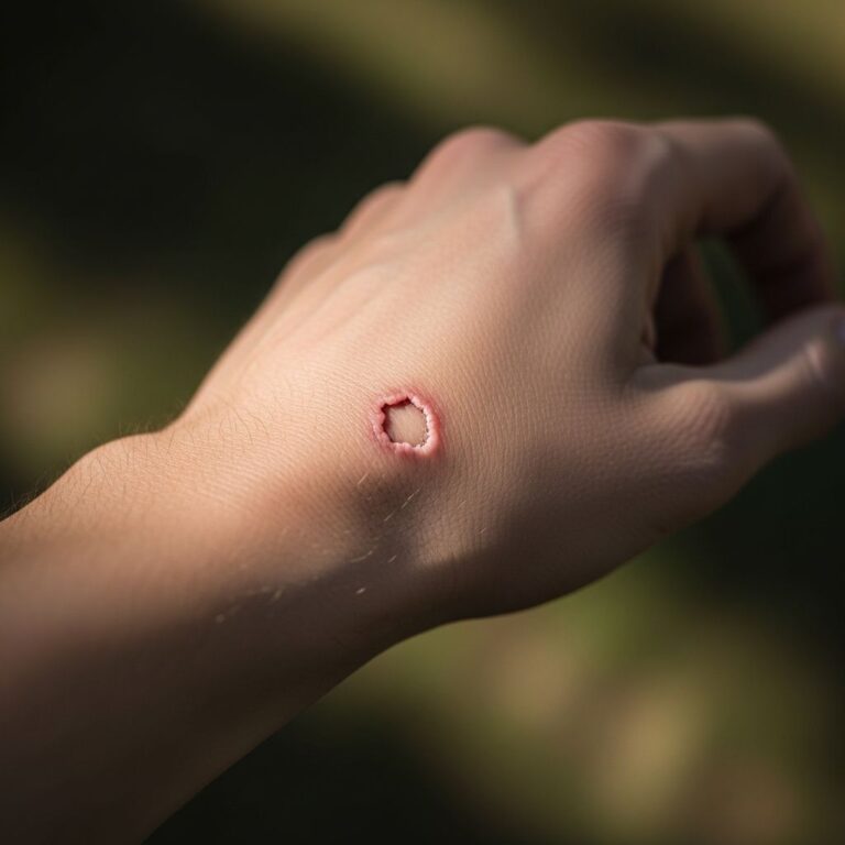
Wound care: The surgical incision requires careful attention during healing. Patients should keep the incision clean and dry, following specific wound care instructions provided by their surgical team. The incision will leave some permanent scarring, and hair may not grow back completely in the scar tissue.
Risks and Complications of Craniotomy
Like all surgical procedures, craniotomy carries potential risks and complications. The specific risks for each patient depend on the location of the brain being operated on, the complexity of the procedure, the patient’s age, and their overall health status.
Common neurological risks include:
– Speech difficulties or language problems
– Temporary or permanent paralysis or weakness
– Vision changes or visual field defects
– Memory loss or cognitive changes
– Balance and coordination difficulties
– Coma or altered consciousness
Other potential complications include:
– Infection at the surgical site or within the brain
– Blood clots
– Bleeding or hemorrhage
– Brain swelling (cerebral edema)
– Stroke
– Seizures
– Anesthesia-related complications
While many of these complications are temporary and resolve with appropriate treatment, some can be permanent. The neurosurgery team works with each patient to discuss these risks thoroughly before surgery and to develop strategies to minimize complications. Patients should not hesitate to contact their healthcare provider if they experience concerning symptoms during recovery, such as severe headache, vision changes, weakness, difficulty speaking, or confusion.
Advanced Craniotomy Techniques
Modern neurosurgery continues to evolve with new techniques designed to minimize risk to the brain and surrounding structures. Johns Hopkins Medicine has developed innovative approaches such as pericranial-onlay cranioplasty, a technique that minimizes risk to the underlying brain and its protective coverings by creating a new surgical corridor rather than reopening the previous scar. This approach keeps the brain protected throughout the entire reconstruction procedure, reducing potential complications.
Awake craniotomy is another advanced technique where patients remain conscious during portions of the surgery. This allows neurosurgeons to directly test brain function and identify critical areas, ensuring maximal tumor removal while preserving essential functions like speech and motor control.
Frequently Asked Questions
Q: How long does a craniotomy procedure take?
A: The surgical procedure typically lasts several hours, depending on the complexity of the brain pathology and the extent of surgery required. The surgical team will provide specific time estimates based on the individual patient’s condition.
Q: Will I have permanent scarring after craniotomy?
A: Yes, the surgical incision will leave permanent scarring on the scalp. Additionally, hair may not grow back completely in the scar tissue. However, depending on the incision location, the scar may be partially hidden by hair as it regrows.
Q: What is the success rate of craniotomy?
A: Success rates vary significantly depending on the specific condition being treated, the patient’s age and health status, and the surgeon’s experience. Your neurosurgeon can provide individualized information based on your particular diagnosis.
Q: Can I return to normal activities after craniotomy?
A: Most patients gradually return to normal activities, though the timeline varies. Physical activity should be gradually increased under medical guidance. Some patients may experience lasting effects on certain functions and may require ongoing rehabilitation or therapy.
Q: Are there alternatives to craniotomy?
A: Depending on the diagnosis, alternative treatments may include radiation therapy, chemotherapy, or stereotactic radiosurgery. Your neurosurgery team will discuss all available options and recommend the approach most appropriate for your condition.
Q: How often is craniotomy performed?
A: Craniotomy is one of the most commonly performed neurosurgical procedures. Hospitals and specialized neurosurgery centers perform thousands of craniotomies annually across the United States.
References
- Craniotomy: What is it, and is it different from craniectomy? — Medical News Today. 2023. https://www.medicalnewstoday.com/articles/craniotomy
- What is a Craniotomy and What is it Used to Treat? — Rebound MD. 2023. https://www.reboundmd.com/news/what-craniotomy-what-it-used-treat
- Johns Hopkins Multidisciplinary Adult Cranioplasty Center | Q&A — Johns Hopkins Medicine. https://www.youtube.com/watch?v=hj4-4PndWOc
Similar Articles


Read full bio of Sneha Tete