Cutaneous Larva Migrans: Parasitic Infection Guide
Understand cutaneous larva migrans: causes, symptoms, diagnosis, and effective treatment options.

Cutaneous Larva Migrans: Understanding the Parasitic Skin Infection
Cutaneous larva migrans (CLM) is a parasitic skin infection caused by the migration of hookworm larvae through the epidermis, resulting in distinctive creeping tracks and intense itching. This dermatological condition occurs when parasitic nematode larvae penetrate exposed skin and migrate through the upper layers, creating characteristic serpentine pathways that resemble winding lines or maps on the skin.
The infection is most commonly encountered in tropical and subtropical geographic locations, including the southeast United States, Latin America, Southeast Asia, and Africa. While the condition is generally self-limiting and resolves without treatment within 4 to 8 weeks, prompt medical intervention can significantly reduce symptoms and prevent secondary bacterial infections resulting from excessive scratching.
Etiology and Causative Organisms
Cutaneous larva migrans is caused by various species of hookworm parasites that normally infect animals rather than humans. The primary causative organisms include:
- Ancylostoma braziliense — the most common cause globally
- Ancylostoma caninum — found in dogs and cats
- Uncinaria stenocephala — occurs in northern regions
- Ancylostoma duodenale — human hookworm occasionally causing CLM
- Necator americanus — human hookworm that rarely causes CLM
These parasites are zoonotic, meaning they naturally infect animals rather than humans, making humans accidental and “dead-end” hosts where the larvae cannot complete their life cycle. The larvae cannot penetrate the basement membrane to reach the gastrointestinal tract, ultimately dying within the skin.
Transmission and Risk Factors
Transmission of cutaneous larva migrans occurs through direct contact of exposed skin with contaminated soil or sand containing parasitic larvae. The infection pathway begins when infected animals deposit feces containing hookworm eggs in warm, moist soil. These eggs hatch into larvae that remain viable in the environment and penetrate human skin upon contact.
Groups at highest risk for cutaneous larva migrans include individuals whose occupations or hobbies involve contact with warm, moist, sandy soil. Specific risk populations comprise:
- Beach visitors and swimmers
- Construction workers and laborers
- Gardeners and agricultural workers
- Military personnel training in endemic areas
- Children playing in contaminated soil
- Individuals walking barefoot in tropical environments
The disease affects people of all ages, sexes, and races who have been exposed to hookworm larvae in endemic regions.
Clinical Presentation and Symptoms
The clinical presentation of cutaneous larva migrans develops over days to months, with a mean time to onset of 5 to 15 days following exposure. The incubation period ranges from 7 days up to 7 months depending on the parasite species and individual factors.
Primary symptoms of cutaneous larva migrans include:
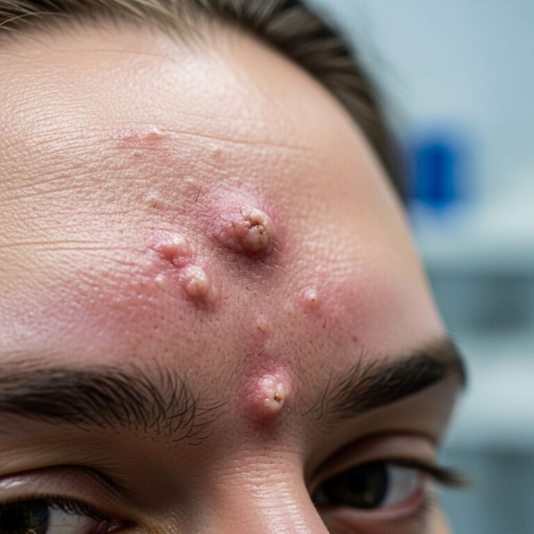
- Initial lesion — a small red dot or bump at the site of penetration
- Intense pruritus — itching that often worsens at night
- Characteristic tracks — red, winding, or wavy lines creating serpentine patterns resembling maps on the skin
- Progressive migration — tracks grow approximately 1 to 2 centimeters daily as the larva moves through the epidermis
- Localized swelling — edema in the affected area
- Secondary bacterial infection — may result from frequent scratching and skin trauma
The most commonly affected sites are the feet, spaces between the toes, hands, knees, and buttocks. While any exposed area can be affected, the feet represent the most frequent location due to increased contact with contaminated soil.
The intense itching frequently interferes with sleep quality and daily activities. Secondary bacterial skin infections represent a significant complication that can develop from repeated scratching, potentially requiring additional antibiotic therapy.
Diagnosis and Clinical Evaluation
Diagnosis of cutaneous larva migrans relies primarily on clinical presentation and patient history rather than laboratory tests. Healthcare providers evaluate:
- The distinctive serpentine or map-like rash pattern
- Recent travel to or residence in tropical or subtropical regions
- History of skin contact with warm, moist sand or soil
- Progressive migration of the lesion over time
- Temporal relationship between exposure and rash onset
The characteristic creeping migration of the rash and associated intense pruritus provide strong diagnostic clues. Dermatological examination reveals the pathognomonic serpentine tracks, which serve as the hallmark diagnostic feature. Biopsy is rarely necessary but may be performed in atypical presentations to exclude other dermatological conditions such as athlete’s foot, thrombophlebitis, or cellulitis.
Natural History and Disease Progression
Cutaneous larva migrans is fundamentally self-limited in humans. The parasitic larvae cannot penetrate the basement membrane of the dermis or migrate to deeper tissues in most cases, preventing access to the gastrointestinal tract where they would complete their life cycle. Consequently, the larvae eventually die from the host’s immune response and physical barriers.
The natural duration of disease varies considerably depending on the hookworm species involved. In most untreated cases, lesions resolve spontaneously within 4 to 8 weeks. However, migration may occasionally continue for several months, and during this extended period, the intense pruritus can severely impact sleep quality and quality of life. In rare cases, the infection may persist for up to one year without treatment.
Treatment Options and Management
Although cutaneous larva migrans resolves without intervention, effective treatment is available to shorten disease duration, reduce symptoms, and prevent secondary bacterial infections. Treatment approaches vary based on the extent of infection and individual patient factors.
Topical Treatment
Topical thiabendazole is considered the treatment of choice for early, localized lesions. The 10% solution or 15% ointment is applied 2 to 3 times daily for 5 to 10 days. Small clinical studies demonstrate that pruritus improvement may occur as early as 48 hours after beginning treatment, with cure rates as high as 98% within ten days.
Advantages of topical therapy include:
- Minimal systemic absorption and side effects
- High efficacy for localized disease
- Direct application to affected areas
Limitations of topical therapy include the requirement for multiple daily applications and reduced utility when multiple lesions are present. Cryotherapy using liquid nitrogen, solid carbon dioxide, or ethylene chloride spray has been historically used but is largely ineffective and should be avoided.
Systemic Treatment
For multiple lesions or severe infestation, oral systemic therapies represent first-line treatment options. Two primary medications demonstrate exceptional efficacy:
Ivermectin is the preferred first-line systemic therapy, requiring only a one-time oral dose of 200 micrograms per kilogram body weight (maximum 12 mg) or one-time dosing for 1 to 2 days. Clinical studies demonstrate cure rates approaching 100% with ivermectin administration. The primary advantage is simplified dosing requiring only one or two doses, improving patient compliance.
Albendazole is another highly effective oral medication administered at 400 mg once daily with a fatty meal for better absorption. Treatment duration ranges from 3 to 5 days, with some studies suggesting a 7-day course may decrease recurrence rates. Cure rates with albendazole approach 100% effectiveness. Mebendazole serves as a second-line alternative depending on medication availability and patient age.
Side effects of systemic therapy are generally mild and may include nausea, vomiting, diarrhea, anorexia, and occasionally a Mazzotti reaction characterized by urticaria, facial swelling, fevers, tachycardia, and tender lymphadenitis.
Supportive Care
Adjunctive measures can help manage symptoms while antiparasitic treatment takes effect:
- Ice packs to temporarily reduce itching and swelling
- Antihistamines to manage pruritus
- Emollients to protect affected skin
- Avoiding scratching to prevent secondary bacterial infection
Completion of the full prescribed course of treatment is essential to ensure complete parasite eradication. Symptoms generally begin to improve within 2 to 3 days of starting medication.
Treatment Comparison Table
| Medication | Dosage | Duration | Cure Rate | Administration |
|---|---|---|---|---|
| Ivermectin | 200 mcg/kg (max 12 mg) | 1-2 days | 94-100% | Oral, once or twice |
| Albendazole | 400 mg daily | 3-7 days | ~100% | Oral with fatty meal |
| Thiabendazole | 10% solution or 15% ointment | 5-10 days | ~98% | Topical, 2-3 times daily |
Prognosis and Outcomes
The prognosis for cutaneous larva migrans is excellent. Disease resolution without treatment represents the rule rather than the exception, as humans cannot serve as definitive hosts for these parasites. Both topical and systemic treatments result in cure rates near 100%, with rapid symptom resolution.
Although recurrence can occur, it is uncommon and responds well to systemic therapy when it develops. Long-term complications are rare in treated cases. The primary concern during the acute infection period is prevention of secondary bacterial infection through symptom management and patient education regarding scratching avoidance.
Prevention and Public Health Measures
Prevention of cutaneous larva migrans focuses on reducing exposure to contaminated soil and sand in endemic regions. Recommended preventive measures include:
- Wearing protective footwear when walking in warm, moist sandy soil
- Avoiding direct skin contact with potentially contaminated soil
- Using barriers such as towels when sitting on beaches
- Avoiding swimming in areas with potential contamination
- Controlling animal populations that may serve as parasite reservoirs
- Implementing public health education in endemic regions
Frequently Asked Questions
Q: How quickly do symptoms develop after exposure to cutaneous larva migrans?
A: Symptoms typically appear within 5 to 15 days after exposure, though the incubation period can range from 7 days to 7 months depending on the parasite species.
Q: Will cutaneous larva migrans go away without treatment?
A: Yes, cutaneous larva migrans is self-limiting and typically resolves within 4 to 8 weeks without treatment. However, treatment significantly reduces symptoms and prevents complications from scratching.
Q: Is cutaneous larva migrans contagious from person to person?
A: No, cutaneous larva migrans cannot be transmitted from person to person. Infection occurs only through direct contact with contaminated soil containing parasitic larvae.
Q: What is the most effective treatment for cutaneous larva migrans?
A: Ivermectin is considered the most effective systemic treatment, offering a one-time or two-dose regimen with cure rates approaching 100%. Topical thiabendazole is preferred for localized early lesions.
Q: Can cutaneous larva migrans affect deeper skin layers or internal organs?
A: In humans, hookworm larvae typically cannot penetrate beyond the dermis due to the basement membrane barrier. Rare cases of larvae reaching deeper tissues such as lungs or intestines have been documented, but this is exceptionally uncommon.
Q: How long does it take to see improvement with treatment?
A: Pruritus improvement may begin as early as 48 hours after starting treatment, with most patients experiencing significant symptom relief within 2 to 3 days.
References
- Cutaneous Larva Migrans: Symptoms, Transmission & Treatment — Tua Saúde. 2024. https://www.tuasaude.com/en/cutaneous-larva-migrans/
- Cutaneous Larva Migrans – StatPearls — National Center for Biotechnology Information (NCBI), National Library of Medicine. 2024. https://www.ncbi.nlm.nih.gov/books/NBK507706/
- Cutaneous Larva Migrans: The Rash Is Not What You Think — Emergency Medicine Residents Association (EMRA). 2024. https://www.emra.org/emresident/article/cutaneous-larva-migrans-case-report
- Cutaneous Larva Migrans – Skin Disorders — Merck Manuals. 2024. https://www.merckmanuals.com/home/skin-disorders/parasitic-skin-infections/cutaneous-larva-migrans
- Cutaneous larva migrans — DermNet New Zealand. 2024. https://dermnetnz.org/topics/cutaneous-larva-migrans
- Clinical Features of Zoonotic Hookworm — Centers for Disease Control and Prevention (CDC). 2024. https://www.cdc.gov/zoonotic-hookworm/hcp/clinical-features/index.html
- Larva Migrans — Center for Food Security and Public Health, Iowa State University. 2024. https://www.cfsph.iastate.edu/Factsheets/pdfs/larva_migrans.pdf
Similar Articles


Read full bio of Sneha Tete