Cutaneous Myiasis: Causes, Symptoms, and Treatment
Understanding cutaneous myiasis: fly larva infestation of the skin and effective treatment options.

Introduction to Cutaneous Myiasis
Cutaneous myiasis is a parasitic skin infection caused by the larvae (maggots) of fly species belonging to the arthropod order Diptera, which consists of two-winged adult flies. The condition occurs when fly larvae infest and feed on the host’s dead or living tissue, body substances, or ingested food. Myiasis affecting the skin represents the most frequently encountered clinical form of myiasis and is the fourth most common travel-associated skin disease.
Myiasis can be categorized clinically based on the area of the body infested, including cutaneous, ophthalmic, auricular, urogenital, and other organ-specific presentations. Cutaneous presentations specifically include three main types: furuncular myiasis, migratory (creeping) myiasis, and wound (traumatic) myiasis, each with distinct clinical characteristics and causative organisms.
Types of Cutaneous Myiasis
Furuncular Myiasis
Furuncular myiasis is characterized by the formation of nodular lesions with a central opening where a single larva develops. The larva creates a cavity connected to the entry point without distant movement, remaining in one location throughout its developmental cycle. This type represents the most widely reported form of cutaneous myiasis globally.
The causative organisms include several important fly species. Dermatobia hominis is the primary cause of furuncular myiasis in Central and South America, while Cordylobia anthropophaga predominates in Africa. Additional causative species include Wohlfahrtia vigil and various Cuterebra species.
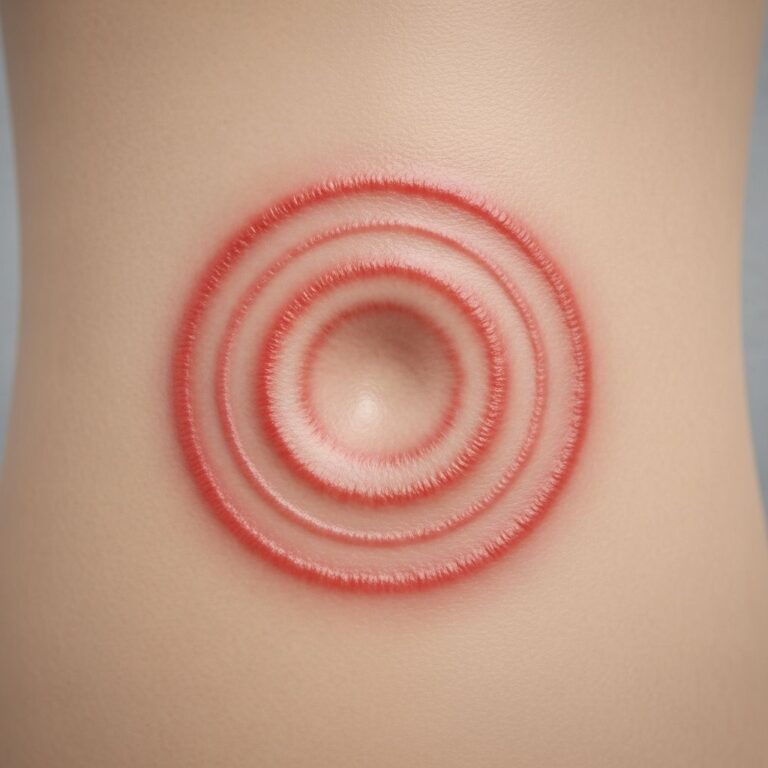
Both Dermatobia hominis and Cordylobia anthropophaga are characterized by their larval stage, which exhibits radial spines on the anterior segments and a spiracle that allows respiration on the posterior segment. The typical clinical presentation consists of an inflammatory nodule approximately 2 centimeters in diameter with a central opening located at the site of egg inoculation. Patients frequently experience itching and a sensation of movement within the lesion.
Clinical patterns vary between species. Dermatobia typically presents as a single lesion associated with an arthropod bite that served as a vector for eggs on exposed skin areas such as limbs or the head, while Cordylobia usually presents as multiple lesions in unexposed areas such as the back or buttocks. Although lesions are generally solitary and occupied by a single larva, cases of multiple simultaneous infestations have been documented.
Migratory Myiasis
Migratory or progressive cutaneous myiasis occurs due to the migration of larvae under the skin surface, creating characteristic creeping patterns. This type differs fundamentally from furuncular myiasis in that the larva does not remain stationary but instead travels through subcutaneous tissues.
The primary fly species responsible for migratory myiasis are Gasterophilus and Hypoderma. These larvae create visible tracks as they migrate through the dermis and subcutis, producing characteristic linear or serpentine lesions that reflect the larval movement patterns.
Wound Myiasis
Wound myiasis occurs when fly larvae infest open wounds in living hosts, parasitizing exposed wound surfaces. Mucous membranes, including oral, nasal, and vaginal membranes, along with body cavity openings such as those in or around the ears and eye sockets, can also be affected.
Primary causative agents include screwworm flies such as Cochliomyia hominivorax and Chrysomya bezziana, as well as Wohlfahrtia magnifica. These species are highly destructive and capable of causing significant tissue damage when they infest wound sites.
Severe cases of wound myiasis may be accompanied by fever, chills, pain, and bleeding from the infested site, with secondary infections occurring frequently. In extreme cases, massive tissue destruction, loss of eyes and ears, erosion of bones and nasal sinuses, and even death can occur. Risk factors for wound myiasis include poor social conditions, poor hygiene, advanced or very young age, psychiatric illness, alcoholism, diabetes, peripheral vascular disease, poor dental hygiene, and physical disabilities that restrict the ability to discourage flies.
Clinical Presentation and Symptoms
The clinical presentation of cutaneous myiasis varies depending on the type and stage of infestation. Typical symptoms include itching, movement beneath the skin, sharp stabbing pain, and a small opening for the larvae to breathe located in nodular redness of the skin.
In furuncular myiasis, patients may experience a sensation of movement within the lesion, which can be quite disturbing. The lesion often appears as an inflammatory nodule with a visible central opening that may occasionally be less obvious if covered by a crust of necrotic debris and larval waste products.
Migratory myiasis produces characteristic serpentine or linear lesions that reflect the path of larval migration through the skin, often accompanied by intense pruritus and a palpable sensation of movement.
Wound myiasis may present with more systemic symptoms, including fever and chills, particularly when secondary bacterial infections develop. Bleeding from the affected site and severe pain are common in this type of myiasis.
Diagnosis of Cutaneous Myiasis
Diagnosis requires a high degree of clinical suspicion based on history-taking and physical examination. The condition should be suspected in patients presenting with a furuncular lesion, particularly if a central opening is observed, and recent travel to tropical areas of Latin America in the case of Dermatobia hominis infestation.
Several diagnostic techniques may be employed:
- Clinical evaluation: Careful examination of the lesion characteristics and patient history regarding recent travel to endemic areas or exposure to contaminated environments
- Dermoscopy: This technique allows visualization of the larva’s posterior spiracles when it is close to the surface, providing direct visualization of the causative organism
- Oxygen deprivation technique: Keeping the dermatoscope on the lesion for several minutes can force the larva to move toward the surface by depriving it of oxygen
- Petroleum jelly occlusion: A simple and cost-effective diagnostic technique involves occluding the opening of the larval channel with petroleum jelly for at least 30 minutes, which reduces oxygen supply to the larva. After this occlusion period, petroleum jelly is removed, and the larva is observed ascending to the surface to breathe
- Imaging studies: Ultrasound can detect a “foreign body” within the lesion, supporting clinical diagnosis
- Specimen identification: Proper identification of the causative agent requires studying anatomical characteristics of the complete specimen, including shape, spines, spiracle, and pigmentation, by an experienced microbiologist or entomologist
The petroleum jelly occlusion technique is particularly valuable because it provides both diagnostic and therapeutic benefits. By forcing the larva to emerge, clinicians can confirm the diagnosis while simultaneously facilitating larval removal.
Treatment Approaches
Treatment of cutaneous myiasis primarily consists of extracting the larva, which may require surgical intervention. Several treatment modalities are available:
- Occlusion method: Occlusion of the opening with waterproof plaster or petroleum jelly can be attempted to make larvae emerge
- Manual removal: Physical extraction of the larva following appropriate preparation and larval mobilization
- Surgical extraction: In some cases, surgical enlargement of the exit opening is necessary to allow complete removal of the specimen without fragmentation
- Larvicides: Chemical agents may be used to kill larvae, though mechanical removal is generally preferred
- Antibiotic therapy: Used to manage secondary bacterial infections, which can occur if dead larvae are not promptly removed
Manual pressure around the lesion is often insufficient for extraction purposes in Dermatobia hominis infestation. Physical occlusion of the exit opening is preferred to force the larva out, although care must be taken to avoid suffocating the specimen inside the lesion, which could lead to an inflammatory response and formation of foreign body granuloma if not removed.
Dead larvae can cause severe inflammatory reactions, making prompt removal essential to minimize tissue damage and complications.
Prevention Strategies
Prevention of cutaneous myiasis is primarily achieved through avoiding exposure to fly species in endemic areas and practicing good wound care and hygiene:
- Avoid traveling to or exercising caution when traveling to endemic regions, particularly Central and South America for Dermatobia hominis and Africa for Cordylobia anthropophaga
- Maintain proper wound hygiene and promptly treat any open wounds to prevent fly infestation
- Use protective clothing and insect repellents when in endemic areas
- Avoid contact with potentially contaminated objects or arthropods that may serve as vectors for eggs
- Practice good personal hygiene and maintain clean living environments
- For individuals at higher risk (elderly, diabetic, immunocompromised), extra precautions should be taken to protect against insect exposure
Pathological Findings
In myiasis, histopathological examination shows the larva of the fly present in the dermis or subcutis with surrounding infiltrate. These pathological findings confirm the parasitic nature of the condition and may assist in definitive diagnosis when clinical assessment is uncertain.
Geographic Distribution and Travel Considerations
Cutaneous myiasis occurs worldwide and represents a significant travel-associated disease. The botfly, originally endemic to Central and South America, along with myiasis-causing species, has become indigenous to parts of Europe due to climate change. This geographic expansion means that clinicians in non-traditional endemic areas must maintain awareness of cutaneous myiasis in patients with appropriate travel histories.
Egg transmission typically takes place indirectly via other insects or objects, highlighting the importance of understanding transmission patterns when assessing risk.
Frequently Asked Questions
Q: How is cutaneous myiasis transmitted to humans?
A: Transmission occurs when fly larvae contact the skin, often through indirect routes via other insects serving as vectors or contaminated objects. Dermatobia hominis eggs are typically deposited on arthropod vectors that then contact human skin.
Q: What is the difference between furuncular and migratory myiasis?
A: Furuncular myiasis involves a larva that creates a stationary cavity with a central opening, remaining in one location. Migratory myiasis, caused by different fly species, involves larvae that migrate through subcutaneous tissues, creating characteristic serpentine tracks.
Q: How long does cutaneous myiasis last without treatment?
A: The duration varies depending on the fly species and type of myiasis. Some cases may persist for weeks or months if untreated, with progressive enlargement and secondary complications potentially developing.
Q: Is cutaneous myiasis dangerous?
A: While uncomplicated cutaneous myiasis is generally not life-threatening, wound myiasis can cause severe complications including massive tissue destruction, secondary infections, systemic symptoms, and rarely, death if left untreated.
Q: Can cutaneous myiasis be treated at home?
A: While petroleum jelly occlusion can be attempted at home as a preliminary measure, proper diagnosis and medical evaluation are essential. Medical professionals may need to surgically enlarge the opening for complete larval removal, particularly with Dermatobia hominis.
Q: What should I do if I suspect I have cutaneous myiasis?
A: Seek immediate medical evaluation, particularly if you have recently traveled to endemic areas. Provide your healthcare provider with a detailed travel history, as this aids diagnosis. Avoid self-treatment with unproven remedies and allow medical professionals to properly identify and remove the larva.
References
- Cutaneous myiasis: a review of the common types of myiasis — Wiley Online Library. 2010. https://onlinelibrary.wiley.com/doi/10.1111/j.1365-4632.2010.04577.x
- Diagnosis of Cutaneous Myiasis — Actas Dermo-Sifiliográficas. 2024. https://actasdermo.org/en-translated-article-diagnosis-cutaneous-myiasis-articulo-S0001731024008275
- Cutaneous myiasis due to botfly larvae — PubMed Central. 2023. https://pmc.ncbi.nlm.nih.gov/articles/PMC9835699/
- Cutaneous myiasis – DermNet — DermNet New Zealand. 2009. https://dermnetnz.org/topics/cutaneous-myiasis
- Myiasis pathology – DermNet — DermNet New Zealand. https://dermnetnz.org/topics/myiasis-pathology
Similar Articles


Read full bio of medha deb