Cutaneous Pseudolymphoma: 4 Key Subtypes & Treatment Guide
Understanding cutaneous pseudolymphoma: causes, diagnosis, and effective treatment strategies for this lymphoma-mimicking skin condition.

Cutaneous pseudolymphoma refers to a heterogeneous group of benign skin reactions characterised by lymphoid infiltrates that may simulate cutaneous lymphoma both clinically and histologically. These conditions are also known as lymphocytoma cutis, cutaneous lymphoid hyperplasia (CLH), or lymphoproliferative disorders. Unlike malignant lymphomas, cutaneous pseudolymphomas are reactive processes that do not progress to systemic disease, though accurate diagnosis is critical to exclude malignancy.
The term encompasses various subtypes, including T-cell-predominant and B-cell-predominant forms, often triggered by external factors such as drugs, infections, or arthropod bites. Diagnosis relies on clinicopathological correlation, with observation sufficient for some reactive cases. Treatment varies by aetiology, ranging from lesion removal to systemic therapies.
What is cutaneous pseudolymphoma?
Cutaneous pseudolymphoma (CPL) describes benign accumulations of lymphocytes and other inflammatory cells in the skin that resemble low-grade lymphomas. These infiltrates can be nodular, plaque-like, or diffuse, primarily affecting the dermis. The condition arises from reactive hyperplasia rather than neoplastic proliferation, distinguished by polyclonality of lymphoid cells (negative T-cell receptor or immunoglobulin gene rearrangements).
Clinically, lesions present as solitary or multiple erythematous papules, nodules, or plaques, often asymptomatic but occasionally pruritic. Common sites include the face, ears, limbs, and trunk. Histologically, dense lymphoid infiltrates with germinal centres mimic follicular lymphoma or mycosis fungoides, necessitating expert review to avoid misdiagnosis.
Subtypes include:
- Lymphocytoma cutis (Spiegler–Fendt sarcoid): B-cell dominant nodules.
- Lymphoplasmacytic plaque: Rare plaque-type with unknown aetiology.
- Drug-induced pseudolymphoma: Resolves upon drug cessation.
- Arthropod bite reactions: Localised lymphoid hyperplasia.
While typically indolent, facial lesions can impair quality of life, and rare progression to lymphoma warrants vigilance.
Who gets cutaneous pseudolymphoma?
CPL affects individuals across ages but peaks in middle-aged adults, with a mean diagnostic age of 42 years. Females predominate, particularly in idiopathic cases, though aetiology influences demographics. For instance, drug-induced forms occur after variable exposure durations (1 day to 15 years).
- Drug-associated: Variable, often in patients on antihypertensives or anticonvulsants.
- Post-leech therapy: Seen in patients treated for venous insufficiency.
- Idiopathic lymphocytoma cutis: More common in women aged 30–60.
Risk factors include hypersensitivity reactions, vaccinations, and trauma. A case series reported nodules on face and limbs following hirudotherapy. Genetic predisposition is unclear, but polyclonal infiltrates confirm benign nature.
What causes cutaneous pseudolymphoma?
Most cases are reactive, triggered by:
- Drugs: Antihypertensives, antidepressants, gold salts; lesions resolve post-discontinuation (weeks to months).
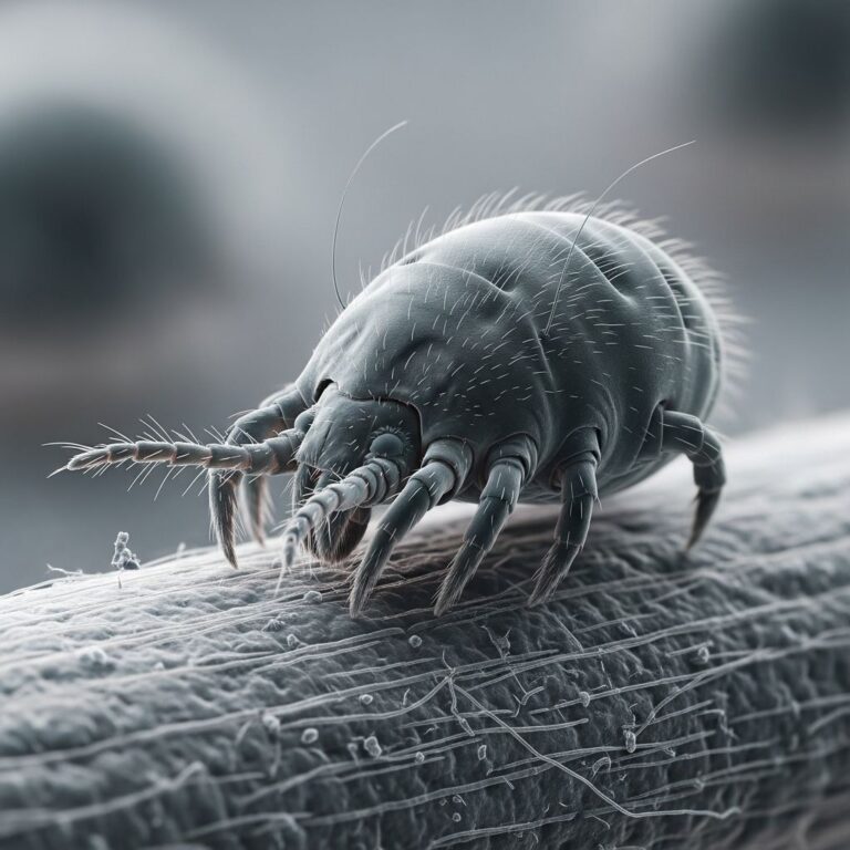
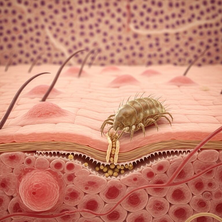
- Arthropod bites/stings: Insect or mite reactions causing localised hyperplasia.
- Infections: Borrelia, herpes viruses, HIV.
- Foreign bodies/trauma: Tattoos, acupuncture, leech therapy.
- Immunisations: Flu vaccine, hypersensitivity reactions.
- Idiopathic: No identifiable trigger, e.g., lymphoplasmacytic plaques.
Pathogenesis involves exaggerated immune responses, with T- or B-cell predominant infiltrates forming germinal centres. In leech-associated cases, salivary antigens provoke nodular reactions.
What are the clinical features of cutaneous pseudolymphoma?
Lesions vary by subtype:
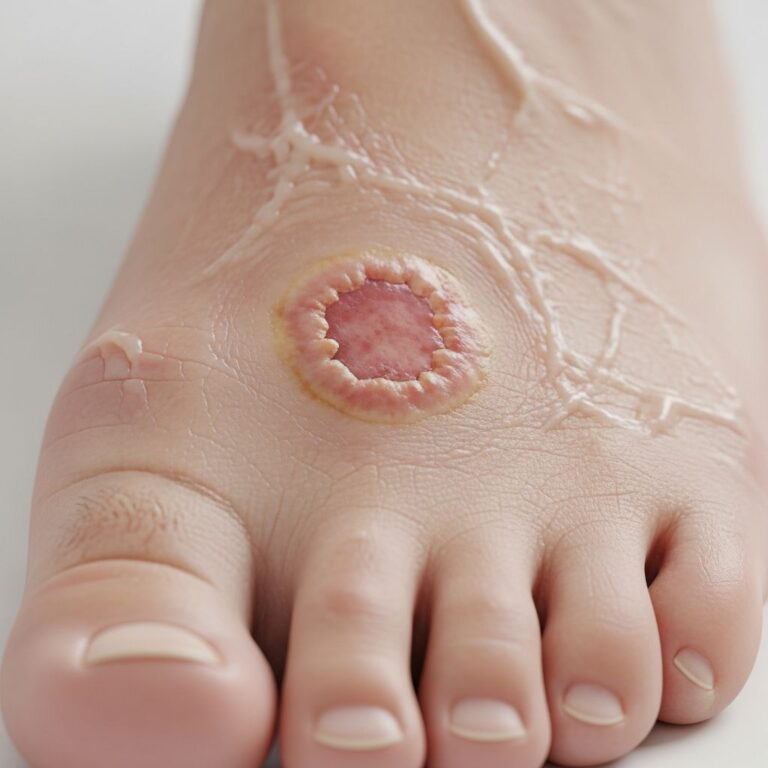
- Solitary or localised: Erythematous to violaceous papules/nodules (0.5–3 cm), firm, on face/ears/limbs.
- Multiple: Grouped papules or plaques mimicking lymphoma.
- Lymphocytoma cutis: Smooth, dome-shaped nodules with telangiectasia.
- Actinic reticuloid: Widespread plaques from photosensitivity.
| Subtype | Typical Presentation | Common Sites |
|---|---|---|
| Lymphocytoma cutis | Single/multiple nodules | Face, ears, nipples |
| Drug-induced | Plaques/nodules | Trunk, limbs |
| Post-leech | Firm red nodules with central depression | Application sites |
| Idiopathic | Plaques or miliary papules | Face, scalp |
Symptoms: Usually asymptomatic; pruritus or burning in 20–30%. Progression is slow; spontaneous regression possible in reactive cases.
How is cutaneous pseudolymphoma diagnosed?
Diagnosis combines clinical features, histology, and molecular studies. Multiple biopsies may be needed due to diagnostic challenges.
- Histology: Dense dermal/periadnexal lymphoid infiltrates; germinal centres in B-cell types; epidermotropism in T-cell.
- Immunohistochemistry: Polyclonal CD4+/CD8+ T-cells or CD20+ B-cells; no light chain restriction.
- Molecular: Polyclonal TCR/IgH gene rearrangements confirm benignity.
- Imaging/Follow-up: Observe behaviour over time.
Differential: Mycosis fungoides, primary cutaneous follicle centre lymphoma, secondary infiltrates. Expert dermatopathology review essential.
What is the treatment for cutaneous pseudolymphoma?
Treatment targets aetiology; observation for indolent cases. A systematic review proposes an algorithm:
- First-line:
- Remove trigger (drugs, allergens).
- Topical/intralesional corticosteroids.
- Second-line: Hydroxychloroquine (400 mg/day, 3 months); PUVA/UVA1.
- Refractory: Rituximab (375 mg/m² weekly x4); thalidomide.
Other options: Imiquimod, excision, PDT. In a case, rituximab resolved facial infiltration lasting 16 months. Leech-induced responded to intralesional steroids + laser, though recurrence noted.
What is the outcome for cutaneous pseudolymphoma?
Excellent prognosis; most resolve with treatment or trigger removal. No progression to lymphoma reported in large series, but long-term follow-up advised. Recurrence possible if trigger persists. Quality of life improves post-treatment, especially facial lesions.
Frequently asked questions
What does cutaneous pseudolymphoma look like?
Red to purple papules, nodules, or plaques, often on face or limbs, mimicking lymphoma.
Is cutaneous pseudolymphoma dangerous?
No, it’s benign, but requires exclusion of true lymphoma.
How is pseudolymphoma treated?
Trigger removal, steroids, antimalarials, or rituximab for refractory cases.
Can pseudolymphoma turn into cancer?
Rarely; polyclonality and observation confirm benign course.
References
- Diagnostic Challenges and Treatment Options for Cutaneous T Cell Pseudolymphoma — PMC/NCBI. 2020-01-15. https://pmc.ncbi.nlm.nih.gov/articles/PMC6977708/
- Treatment of Cutaneous Pseudolymphoma: A Systematic Review — Acta Dermato-Venereologica. 2019-05-08. https://www.medicaljournals.se/acta/content/html/10.2340/00015555-2841
- Cutaneous pseudolymphoma – DermNet — DermNet NZ. 2023. https://dermnetnz.org/topics/cutaneous-pseudolymphoma
- Cutaneous Pseudolymphoma Review — International Society of Dermatopathology. 2021. https://www.intsocdermpath.org/assets/docs/21stJMHandouts/Mitteldorf handout Pseudolymphoma review.pdf
- Cutaneous pseudolymphoma: a case series of three patients — Frontiers in Medicine. 2024-10-11. https://www.frontiersin.org/journals/medicine/articles/10.3389/fmed.2024.1507399/full
Similar Articles


Read full bio of Sneha Tete