Cutaneous T-Cell Lymphoma: Images, Diagnosis & Overview
Visual guide to cutaneous T-cell lymphoma: Diagnostic imaging, clinical features, and treatment approaches.

Cutaneous T-Cell Lymphoma: Overview and Clinical Presentation
Cutaneous T-cell lymphomas (CTCLs) represent a diverse group of non-Hodgkin lymphomas characterized by the malignant proliferation of T lymphocytes in the skin. These conditions encompass a spectrum of indolent to aggressive disorders, with mycosis fungoides and Sézary syndrome representing the most common subtypes. CTCLs are considered the predominant category of primary cutaneous lymphomas, accounting for a significant proportion of skin-based lymphoproliferative disorders.
The pathophysiology of CTCL involves the infiltration of malignant helper/inducer T lymphocytes into the epidermis and dermis, leading to characteristic skin manifestations. Unlike systemic lymphomas, CTCLs typically present with localized skin involvement, though advanced disease may demonstrate extracutaneous spread to lymph nodes and internal organs. The natural history of most CTCLs is indolent, with symptoms potentially present for 2 to 10 years before definitive diagnosis through biopsy confirmation.
Clinical Presentation and Distribution Patterns
CTCL lesions exhibit characteristic distribution patterns that aid in clinical diagnosis. Lesions typically appear in bathing trunk distribution, affecting non-sun-exposed areas such as the buttocks, medial thighs, breasts, and lower abdomen. This distribution pattern distinguishes CTCL from allergic contact dermatitis, which commonly involves the hands, eyelids, face, and perianal regions.
The clinical presentation of CTCL varies depending on disease stage and subtype:
- Patch stage: Early manifestation presenting as erythematous, scaly patches with variable definition. Patches may resemble eczema or other inflammatory dermatoses, leading to diagnostic delays.
- Plaque stage: Progression to infiltrated, well-demarcated plaques that may cover extensive body surface areas. Plaques can become thickened and hyperkeratotic.
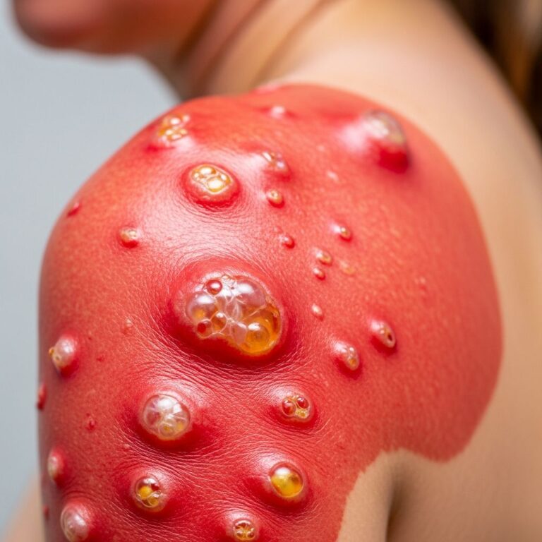
- Tumor stage: Development of nodular lesions representing more aggressive disease. Tumors may ulcerate and become secondarily infected.
- Erythrodermic presentation: Total body erythema with severe pruritus, scaling, and potential hair loss. This presentation indicates advanced systemic disease.
Pruritus represents a significant clinical symptom in many CTCL patients, often preceding visible skin manifestations by months or years. The itching can be severe and refractory to standard therapeutic interventions, substantially impacting quality of life.
Diagnostic Approach and Histopathological Features
Accurate diagnosis of CTCL requires a comprehensive approach combining clinical evaluation, histopathological examination, and ancillary testing. An extensive history and physical examination form the foundation of CTCL diagnosis, with particular emphasis on lesion location, symptom progression, and systemic involvement assessment.
Physical examination should include detailed assessment of:
- Comprehensive skin examination documenting distribution, morphology, and body surface area involvement
- Extensive lymph node palpation of cervical, supraclavicular, axillary, and inguinal regions
- Hepatomegaly and splenomegaly assessment
- Evaluation of potential secondary infections at erosion or ulceration sites
Skin biopsy remains essential for definitive CTCL diagnosis. A broad shave biopsy performed below the dermal-epidermal junction is generally recommended, particularly for early patch and thin plaque lesions. Larger nodular lesions may require deeper sampling to ensure adequate tissue assessment.
Histopathological diagnostic criteria for CTCL include:
- Presence of convoluted (mycosis-like) lymphocytes
- Band-like upper dermal infiltrate
- Epidermal infiltration with Pautrier abscesses (collections of neoplastic lymphocytes)
- Variable degrees of epidermotropism
T-cell receptor (TCR) gene analysis by high-throughput screening identifies clonal T-cell populations, providing molecular evidence supporting malignancy diagnosis. Dominant TCR beta or gamma sequences indicate clonal expansion consistent with lymphoma pathology.
Staging and Prognostic Assessment
CTCL staging employs the TNM classification system designating tumor (skin involvement), node (lymph node involvement), and metastasis (systemic spread). This staging framework guides treatment planning and prognostic assessment.
The International Society for Cutaneous Lymphomas and European Organisation for Research and Treatment of Cancer have proposed a formal staging system incorporating:
- T classification: Ranging from T1 (limited patches/plaques) to T4 (generalized patches, plaques, or tumors with erythroderma)
- N classification: Ranging from N0 (no lymph node involvement) to N3 (involvement of multiple node regions)
- M classification: Indicating absence (M0) or presence (M1) of visceral organ involvement
Peripheral blood involvement with circulating CTCL cells correlates with advanced skin stage, lymph node involvement, visceral disease, and shortened survival, necessitating careful blood assessment in disease evaluation.
Treatment Approaches by Disease Stage
CTCL management is individualized based on disease stage, burden, and patient factors. While curative modalities remain elusive except in minimal skin-confined disease, multiple therapeutic options achieve disease control and symptom palliation.
Early-Stage Disease (Stage I-II)
Early-stage CTCL limited to skin involvement benefits from skin-directed therapies as initial management. These approaches include:
- Topical corticosteroids: High-potency agents represent first-line topical therapy for limited lesions
- Topical mechlorethamine (nitrogen mustard): Second-line topical agent for resistant patches and plaques
- Phototherapy: Narrowband UVB (NB-UVB) for widespread patch and thin plaque disease; PUVA (psoralen plus UVA) or UVA1 for thicker plaques
- Localized radiation therapy: Combined with phototherapy for thicker plaques or nodular lesions
- Photodynamic therapy: Alternative approach for selected patients
- Systemic retinoids and interferons: Adjunctive agents combined with phototherapy for enhanced response
Advanced-Stage Disease (Stage III-IV)
Advanced CTCL with blood involvement, lymph node involvement, or systemic manifestations requires systemic therapeutic approaches:
- Extracorporeal photopheresis (ECP): For patients with blood involvement
- Total skin electron beam therapy (TSEBT): For extensive cutaneous disease, potentially combined with ECP
- Combination chemotherapy: Multiagent regimens including SMILE (dexamethasone, methotrexate, ifosfamide, L-asparaginase, etoposide)
- Hematopoietic stem cell transplantation: Allogeneic transplant for selected patients with aggressive histology or chemotherapy-refractory disease
- Targeted therapy: Novel agents targeting specific molecular pathways
Special Considerations in CTCL Management
Several clinical scenarios require specific management approaches. Erythrodermic presentations demand careful assessment for secondary infections, with empiric anti-staphylococcus aureus coverage often indicated. Gram-negative coverage may be necessary for patients with erosions or ulcerations.
Assessment of erosions should include evaluation for herpes simplex virus and varicella zoster virus infection, particularly in immunocompromised patients. Infectious disease consultation becomes prudent when managing multi-drug resistant cutaneous infections.
Biopsies should be repeated if clinicopathologic discrepancy exists, if patients develop new nodular lesions, or if lymphocyte exocytosis without diagnostic features appears on initial examination. Corticosteroids should be discontinued at least 2 weeks before biopsy to avoid suppression of lymphocytic infiltration potentially obscuring diagnostic findings.
CTCL Subtypes
Beyond mycosis fungoides and Sézary syndrome, other CTCL subtypes require distinct diagnostic approaches:
- Primary cutaneous gamma-delta T-cell lymphoma: Rare aggressive variant requiring chemotherapy or stem cell transplantation
- Primary cutaneous CD8-positive aggressive epidermotropic cytotoxic T-cell lymphoma: Rapidly progressive variant presenting with widespread papules, plaques, and tumors with potential mucosal involvement. Treatment typically requires combination chemotherapy or stem cell transplantation.
- Primary cutaneous T-cell lymphoma not otherwise specified: Heterogeneous group requiring individualized treatment based on clinical and pathological features
Diagnostic Pitfalls and Challenging Cases
Diagnosis of early CTCL presents significant challenges, as patch-stage disease may closely resemble benign inflammatory dermatoses such as atopic dermatitis, psoriasis, or contact dermatitis. Symptoms may persist for years before diagnostic confirmation, necessitating low threshold for repeat biopsy in clinically suspicious cases.
Recurrent dermatitis after patch testing and allergen avoidance, or dermatitis refractory to standard therapeutic interventions, should prompt re-biopsy consideration. Similarly, chronic residual dermatitis despite appropriate treatment warrants histological re-evaluation to exclude evolving CTCL.
Biopsy tissue quality significantly impacts diagnostic accuracy. Adequate sampling through broad shave biopsies provides superior diagnostic yield compared to punch biopsies, particularly for early-stage disease. Multiple biopsies from different lesional sites may be necessary to confirm clonal T-cell populations through TCR analysis.
Prognosis and Long-Term Outcomes
Most CTCLs follow an indolent clinical course, with many patients experiencing long survival even without curative treatment. However, disease progression may occur, transitioning from patch to plaque to tumor stages over years or decades. Blood involvement and visceral disease indicate more aggressive biology and worse prognosis.
Factors influencing prognosis include disease stage at diagnosis, presence of blood involvement, lymph node involvement patterns, and histological subtype. Early diagnosis and aggressive management of limited-stage disease may prolong disease-free intervals and optimize quality of life.
Frequently Asked Questions
Q: Is cutaneous T-cell lymphoma curable?
A: Most CTCLs are treatable but not curable, with the possible exception of minimal disease confined to the skin. Treatment focuses on disease control and symptom palliation rather than definitive cure.
Q: How long does it take to diagnose CTCL?
A: Diagnosis may be delayed years from symptom onset, as early patch-stage disease resembles benign dermatoses. Symptoms typically persist 2 to 10 years before biopsy confirmation.
Q: Can early-stage CTCL be managed with topical therapy alone?
A: Yes, early-stage disease limited to skin involvement often responds well to skin-directed therapies including topical corticosteroids, topical mechlorethamine, and phototherapy.
Q: What does erythroderma indicate in CTCL patients?
A: Erythroderma represents advanced disease with total body skin involvement and potential blood involvement, requiring systemic therapeutic approaches beyond skin-directed therapies.
Q: How often should patients with CTCL be monitored?
A: Regular clinical monitoring with dermatological assessment, lymph node examination, and appropriate staging studies should occur based on disease stage and treatment response, typically at intervals determined by disease activity.
References
- A Clinician’s Guide to Cutaneous T-Cell Lymphoma Presenting as Atypical Dermatitis — National Center for Biotechnology Information (NCBI/PMC). Accessed January 2026. https://pmc.ncbi.nlm.nih.gov/articles/PMC8484943/
- Cutaneous Lymphoma – A Patient’s Guide — Lymphoma Coalition. 2020. https://lymphomacoalition.org/lymphoma-types/cutaneous-lymphoma/
- Mycosis Fungoides and Other Cutaneous T-Cell Lymphomas Treatment — National Cancer Institute (Cancer.gov). Accessed January 2026. https://www.cancer.gov/types/lymphoma/hp/mycosis-fungoides-treatment-pdq
- Cutaneous T-Cell Lymphomas (CTCL) — Merck Manuals Professional Version. Accessed January 2026. https://www.merckmanuals.com/professional/hematology-and-oncology/lymphomas/cutaneous-t-cell-lymphomas-ctcl
- A Patient’s Guide to Understanding Cutaneous Lymphoma — Cutaneous Lymphoma Foundation. 2018. https://www.clfoundation.org/patient-education
- Cutaneous T-Cell Lymphomas (CTCL) Treatment Algorithm — MD Anderson Cancer Center. 2023. https://www.mdanderson.org/for-physicians.html
Similar Articles


Read full bio of Sneha Tete