Cysticercosis: Causes, Symptoms, Diagnosis & Treatment
Understanding cysticercosis: A parasitic infection caused by tapeworm larvae requiring prompt medical attention.

Cysticercosis: A Comprehensive Overview
Cysticercosis is an infection caused by the larvae of the pork tapeworm, scientifically known as Taenia solium. This parasitic condition develops when a person ingests microscopic tapworm eggs, typically through contaminated food or water. Once inside the body, these eggs hatch and release larvae that migrate through the bloodstream to various tissues, including muscle, brain, eye, and other organs, where they form cyst-like structures called cysticerci.
When cysts develop in the brain or central nervous system, the condition is specifically referred to as neurocysticercosis. This variant represents one of the most serious forms of the infection and can lead to significant neurological complications. While cysticercosis can affect various body systems, the neurological manifestations are often the most concerning and require immediate medical intervention.
How Cysticercosis Develops
Understanding the transmission pathway of cysticercosis is crucial for prevention and early detection. The infection begins when a person consumes food or water contaminated with eggs from the pork tapeworm Taenia solium. Unlike taeniasis (which occurs from eating undercooked pork containing cysts), cysticercosis stems specifically from ingesting the eggs of the parasite.
After ingestion, the microscopic eggs travel to the small intestine where they hatch. The released larvae penetrate the intestinal wall and enter the bloodstream. From there, they can circulate throughout the body and lodge in various tissues. The larvae then encyst themselves, forming fluid-filled sacs that contain the parasitic organism. This encystation process can take weeks to months, and the cysts may remain dormant in tissues for extended periods.
Poor sanitation and hygiene practices significantly increase transmission risk, particularly in areas where infected individuals have inadequate sanitation facilities. Regions with high prevalence include Central America, South America, sub-Saharan Africa, India, and Asia.
Signs and Symptoms of Cysticercosis
The clinical presentation of cysticercosis varies considerably depending on the number of cysts present and their anatomical locations. Many infected individuals remain asymptomatic for months or even years after initial infection. Symptoms typically emerge when cysts begin degenerating and dying, triggering inflammatory responses in surrounding tissues.
Common symptoms of cysticercosis include:
- Seizures (particularly common in neurocysticercosis)
- Chronic headaches lasting weeks or months
- Vision problems and potential blindness if eye involvement occurs
- Memory difficulties and cognitive changes
- Muscle pain and weakness
- Lumps beneath the skin in muscle areas
- Fever and general malaise
In cases of neurocysticercosis, symptoms can be particularly severe. Brain edema (swelling) frequently accompanies cyst degeneration, leading to increased intracranial pressure. Patients may experience focal neurological deficits, visual field loss, photophobia (light sensitivity), and in severe cases, dementia or significant cognitive decline.
The severity of symptoms does not always correlate with the number of cysts present. Sometimes extensive infection produces minimal symptoms, while other cases with fewer cysts cause debilitating manifestations. This unpredictability underscores the importance of medical evaluation for anyone with risk factors presenting with suggestive symptoms.
Diagnostic Approaches for Cysticercosis
Accurate diagnosis of cysticercosis requires a systematic approach combining clinical history, imaging studies, and laboratory testing. Healthcare providers will typically inquire about travel history, dietary habits, and specific symptomatology to establish clinical suspicion.
Imaging Studies
Neuroimaging forms the cornerstone of cysticercosis diagnosis. Magnetic resonance imaging (MRI) and computed tomography (CT) scans are the primary imaging modalities used to identify cysts within brain tissue and other organs. These imaging studies can reveal cysts in different developmental stages:
- Vesicular stage: living larvae within fluid-filled sacs
- Colloidal stage: larvae in early degeneration
- Granulonodular stage: cyst wall thickening with advancing degeneration
- Calcification stage: fully calcified lesions representing resolved infection
Advanced imaging techniques like optical coherence tomography (OCT) can detect cysts in ocular structures, while fluorescein angiography may reveal associated inflammatory changes.
Laboratory Testing
Blood tests detecting cysticercus antibodies support diagnosis and can indicate acute versus chronic infection. However, serological testing has limitations, particularly in patients with single cysts or calcified lesions, where false negatives are more common. Negative test results do not exclude infection, especially in localized disease.
In atypical presentations, healthcare providers may recommend lumbar puncture (spinal tap) to analyze cerebrospinal fluid or brain biopsy when diagnosis remains uncertain. If surgical intervention becomes necessary, pathological examination of extracted cyst material confirms diagnosis definitively.
Specialized Examinations
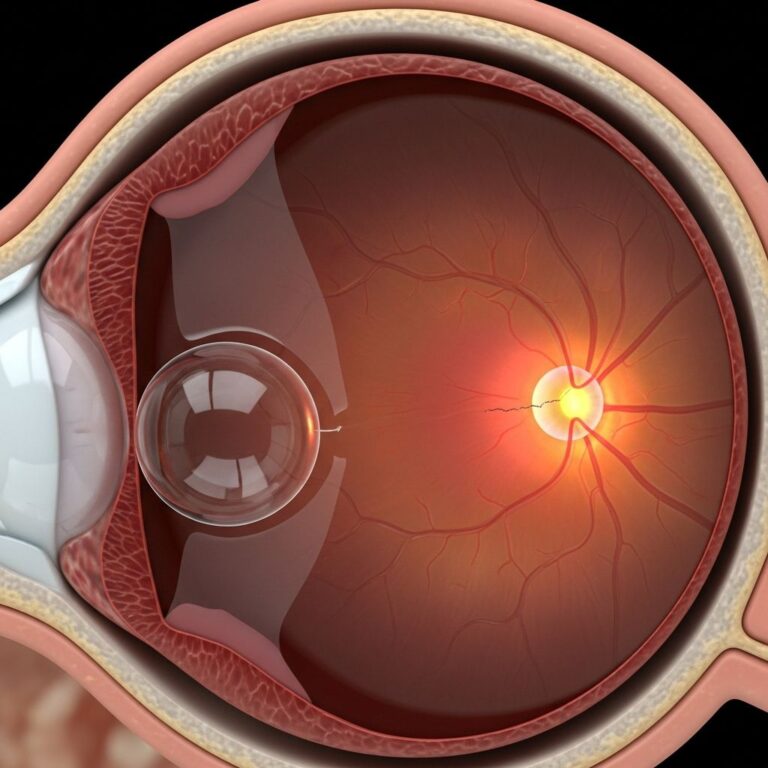
Ophthalmological examination is recommended when cysticercosis affects ocular structures, as parasitic involvement can cause choroiditis (inflammation of the choroid layer) and potentially lead to vision loss.
Treatment Options for Cysticercosis
Treatment approaches for cysticercosis are highly individualized, depending on symptom severity, cyst location, number of lesions, and the patient’s response to medical therapy. Some cases may not require parasite-killing treatment, while others demand aggressive intervention.
Antiparasitic Medications
When warranted, infections are generally treated with antiparasitic drugs such as albendazole or praziquantel. These medications directly target and kill the parasitic larvae within their cysts. Treatment duration typically ranges from 7 to 30 days depending on the specific drug and infection severity.
Anti-inflammatory Agents
Corticosteroids and anti-inflammatory medications accompany antiparasitic therapy to manage inflammation triggered by dying parasites. This inflammation can cause or exacerbate brain swelling, necessitating aggressive anti-inflammatory management to prevent neurological deterioration.
Antiseizure Medications
Many patients require antiseizure medications, either acutely during infection or chronically afterward. Some individuals continue experiencing seizures even after successful parasite eradication due to calcified scar tissue remaining in the brain. Long-term antiseizure therapy may be necessary in these cases.
Surgical Intervention
Surgery becomes necessary in specific circumstances, including cyst location in critical brain regions, insufficient response to medical therapy, significant brain swelling, or when parasites lodge in the eye or other vital organs. Surgical removal provides direct access to problematic lesions while allowing pathological confirmation of diagnosis.
Treatment Outcomes
Many patients begin feeling better within weeks of appropriate treatment and experience no long-term complications. However, severe cases may result in persistent symptoms even after successful parasite elimination. Calcifications (scar-like lesions) can remain indefinitely in the brain, potentially causing continued seizures or other neurological effects.
Prevention Strategies for Cysticercosis
Prevention of cysticercosis relies primarily on maintaining good personal hygiene and avoiding contaminated food sources. Key prevention measures include:
- Practicing thorough handwashing before eating and after bathroom use
- Consuming only clean drinking water from reliable sources
- Properly washing fruits and vegetables before consumption
- Ensuring adequate sanitation facilities in communities
- Avoiding foods prepared in areas with poor hygiene standards
- Being cautious when traveling to endemic regions
- Supporting public health initiatives targeting sanitation improvements
Travelers to high-risk regions should exercise particular vigilance regarding food and water safety.
Long-term Complications and Prognosis
While many individuals recover well after appropriate treatment, some face lasting complications. Post-infection seizures remain a significant concern, occurring in patients even after parasites are eliminated, particularly when brain calcifications develop.
Vision problems represent another potential long-term consequence, especially when parasites affect ocular structures. Cognitive changes and memory impairment may persist in cases involving extensive brain involvement.
The prognosis for cysticercosis improves substantially with early diagnosis and appropriate treatment. Prompt medical intervention can prevent progression to severe neurological complications and reduce the likelihood of permanent disability.
Frequently Asked Questions About Cysticercosis
Q: How long can cysticercosis remain dormant in the body?
A: Cysticercosis cysts can remain dormant and asymptomatic for months to years after infection. Symptoms typically appear when cysts begin degenerating and causing inflammation in surrounding tissues.
Q: Can cysticercosis be fatal?
A: While cysticercosis can cause serious complications, particularly when affecting the brain, modern treatment options have significantly improved outcomes. Fatal cases are relatively uncommon with appropriate medical care, though severe untreated neurocysticercosis can lead to life-threatening complications.
Q: Is cysticercosis contagious between people?
A: Direct person-to-person transmission of cysticercosis does not occur. Infection requires ingestion of tapeworm eggs, typically through contaminated food or water. However, infected individuals with poor sanitation practices can contaminate their environment, putting others at risk.
Q: What regions have the highest cysticercosis prevalence?
A: Cysticercosis is most common in Central America, South America, sub-Saharan Africa, India, and Asia, particularly in areas with inadequate sanitation and hygiene infrastructure.
Q: Can imaging always detect cysticercosis cysts?
A: While MRI and CT scans are highly effective at detecting brain cysts, some early-stage or calcified lesions may be difficult to visualize. Blood tests and clinical history help supplement imaging findings.
Q: Will I need lifelong treatment for cysticercosis?
A: Treatment duration varies considerably. While antiparasitic therapy typically lasts weeks to months, some patients require long-term antiseizure medication to manage complications arising from calcified lesions.
References
- About Cysticercosis — Centers for Disease Control and Prevention. 2024. https://www.cdc.gov/cysticercosis/about/index.html
- Neurocysticercosis: What It Is, Symptoms & Treatment — Cleveland Clinic. 2024. https://my.clevelandclinic.org/health/diseases/neurocysticercosis
- Case Study: Cysticercosis Causes Bilateral Multifocal Choroiditis — Cleveland Clinic. 2024. https://consultqd.clevelandclinic.org/case-study-cysticercosis-causes-bilateral-multifocal-choroiditis
- Worsening migraine due to neurocysticercosis — Cleveland Clinic Journal of Medicine. 2016. https://www.ccjm.org/content/84/3/196
- Cysticercosis/Taeniasis — Texas Department of State Health Services. 2024. https://www.dshs.texas.gov/sites/default/files/IDCU/health/zoonosis/disease/cystipam.pdf
Similar Articles


Read full bio of medha deb