Cystography: Bladder Imaging for Diagnosis
Understanding cystography: A comprehensive guide to bladder imaging and diagnosis procedures.

What is Cystography?
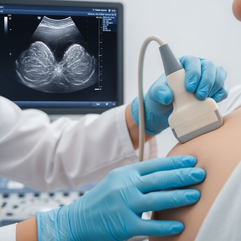
Cystography is a specialized medical imaging procedure that allows healthcare providers to visualize the bladder by using contrast dye and X-ray technology. During this fluoroscopic study, a contrast dye is injected into the bladder through a catheter, and X-rays are then taken to examine the organ in detail. This procedure differs from a voiding cystourethrogram (VCUG) in that cystography focuses specifically on the bladder itself, rather than the posterior urethra.
The primary purpose of cystography is to provide doctors with detailed, clear images of the bladder to help identify various conditions, abnormalities, and potential health concerns. The contrast dye makes the bladder visible on X-ray images, allowing for precise visualization of its structure and function.
Why Cystography is Performed
Your healthcare provider may recommend a cystography procedure if you are experiencing specific symptoms or have conditions that require detailed bladder evaluation. Common reasons for ordering this test include:
- Difficulty beginning or completing urination
- Difficulty emptying the bladder completely
- Urgent or frequent need to urinate
- Urinary incontinence
- Blood in the urine (hematuria)
- Pain or discomfort during urination
- Suspected bladder cancer or other malignancies
- Evaluation of the urinary system after trauma or injury
Cystography is particularly valuable for diagnosing cancers of the bladder, urinary system, and prostate cancer. It can also detect other bladder problems such as stones, strictures, diverticula, and structural abnormalities. In some cases, a biopsy may be performed during cystography if concerning tissue or a tumor is located during the examination.
Preparation for Cystography
Proper preparation is essential for ensuring the procedure is conducted safely and effectively. Your healthcare provider will give you specific instructions tailored to your individual situation. General preparation guidelines typically include:
- Abstaining from solid food for a specified period before the procedure
- Managing your fluid intake as directed by your healthcare team
- Emptying your bladder completely before the procedure begins
- Removing all clothing and wearing a hospital gown
- Informing your doctor of any allergies, especially to iodine or contrast dye
- Notifying your healthcare provider of any medications you are taking
- Arranging for transportation home after the procedure, as you may not be able to drive immediately
It is important to discuss any concerns or questions about preparation with your healthcare provider before your scheduled appointment.
The Cystography Procedure: Step-by-Step
Understanding what to expect during cystography can help reduce anxiety and ensure you are mentally prepared for the examination. Here is a detailed breakdown of the typical procedure:
Initial Positioning and Preparation
You will be asked to lie flat on your back on an X-ray table. Your feet may be positioned in stirrups with your knees bent to provide optimal access for the catheter insertion. The area around the opening of your urethra will be cleaned with a sterile solution and disinfectant. A numbing gel or local anesthetic will be applied to help prevent discomfort during catheter insertion.
Catheter Insertion
Once the area is properly numbed and prepared, a healthcare provider will carefully insert a thin, flexible or rigid urinary catheter into your urethra and guide it into your bladder. This catheter serves as the conduit through which the contrast dye will be introduced into your bladder. You may experience some pressure or mild discomfort during insertion, but the numbing agent should minimize any pain.
Baseline X-Ray
Before injecting the contrast dye, your healthcare provider will take an initial X-ray of your kidney, ureter, and bladder (often called a KUB film) to ensure they can visualize the entire urinary system clearly. For male patients, a lead shield will be positioned over the testes to protect the gonads from radiation exposure.
Contrast Dye Injection
The contrast dye is then injected into your bladder through the catheter. The catheter tubing will be clamped to prevent the dye from draining out of the bladder prematurely. As the bladder fills with the dye, you may experience the sensation of needing to urinate, which is a normal part of the procedure. The dye makes your bladder visible on X-ray images, providing clear, detailed pictures of its structure and any abnormalities.
X-Ray Imaging
X-rays will be taken during the injection of the contrast dye and afterward. You may be asked to change positions multiple times to allow the radiologist to capture different views of your bladder from various angles. These multiple perspectives help ensure that all areas of the bladder are thoroughly evaluated.
Catheter Removal and Post-Imaging
Once all necessary X-ray images have been obtained, the catheter will be carefully removed from your urethra. If a voiding cystography component is included in your procedure, you will be asked to urinate on the X-ray table while additional images are captured. This allows your healthcare provider to see how your bladder functions during emptying and to visualize the direction of urine flow.
The entire procedure typically takes 30 to 45 minutes to complete, though the exact duration may vary depending on your individual circumstances and the complexity of your case.
After the Procedure
Following your cystography procedure, your healthcare provider will provide you with specific aftercare instructions to promote comfort and healing. Important post-procedure guidelines include:
Hydration
You will be advised to drink large amounts of clear fluids for several days following the procedure. This increased fluid intake helps flush the contrast dye from your bladder and urinary system, promoting complete elimination of the dye.
Urinary Changes
Due to the presence of the contrast dye in your system, your urine may appear discolored or have an unusual color for a short time after the procedure. This is completely normal and should resolve as you continue to hydrate and flush your system.
Activity and Recovery
Most patients can return to their normal activities immediately following the procedure. However, you should arrange for someone to drive you home if sedation was used during your examination, as you may not be fit to drive immediately afterward.
Monitoring for Complications
While complications are rare, you should contact your healthcare provider if you experience persistent pain, fever, inability to urinate, or continued hematuria beyond what was present before the procedure.
Risks and Side Effects
Cystography is generally considered a safe procedure with minimal risk. However, as with any medical intervention, there are potential side effects and risks to be aware of:
Common Side Effects
Most patients experience minimal discomfort during and after the procedure. Some may report mild urinary discomfort or a temporary urge to urinate. Discomfort typically resolves within a few days.
Rare Complications
Serious complications are uncommon but may include urinary tract infection, allergic reaction to the contrast dye, urinary retention, and bladder perforation. Radiation exposure is minimal, and patients generally do not experience any long-term side effects from the procedure.
Understanding Your Results
Following your cystography procedure, your healthcare provider will analyze the detailed X-ray images to evaluate your bladder and urinary system. The results will help your doctor assess for various conditions and abnormalities.
What Normal Results Show
Normal cystography results indicate a bladder of appropriate size and shape, with no evidence of growths, stones, strictures, or other abnormalities. The bladder should fill and empty appropriately without obstruction.
What Abnormal Results May Indicate
Abnormal findings may reveal bladder cancer, benign tumors, bladder stones, strictures, diverticula, or other urinary tract abnormalities. Your healthcare provider will discuss the findings with you in detail and recommend appropriate next steps for treatment or further evaluation.
Comparison with Other Diagnostic Procedures
| Procedure | Primary Focus | Imaging Method | Use of Contrast | Duration |
|---|---|---|---|---|
| Cystography | Bladder evaluation | X-ray/Fluoroscopy | Contrast dye injected | 30-45 minutes |
| VCUG (Voiding Cystourethrogram) | Bladder and urethra | X-ray/Fluoroscopy | Contrast dye injected | Less than 1 hour |
| Cystoscopy | Direct bladder visualization | Optical tube with camera | No contrast dye | 30-60 minutes |
| Ultrasound | Bladder assessment | Sound waves | No radiation or contrast | 20-30 minutes |
Frequently Asked Questions
Q: Is cystography painful?
A: Most patients experience minimal pain during cystography. A numbing gel is applied before catheter insertion to reduce discomfort. You may feel pressure or mild discomfort, but the procedure is generally well-tolerated by most patients.
Q: How long does cystography take?
A: The procedure typically takes 30 to 45 minutes to complete. The exact duration depends on your individual circumstances, the complexity of your case, and whether additional imaging views are necessary.
Q: What should I do if I’m allergic to iodine or contrast dye?
A: Inform your healthcare provider immediately if you have a known allergy to iodine or contrast dye. Your doctor may recommend premedication, use of alternative contrast agents, or alternative diagnostic procedures.
Q: Can I eat or drink before cystography?
A: You will typically be instructed to abstain from solid food and manage your fluid intake before the procedure. Your healthcare provider will give you specific fasting instructions based on your individual situation.
Q: When will I receive my results?
A: A radiologist will analyze your X-ray images after the procedure. Your healthcare provider will typically discuss results with you within a few days and recommend any necessary follow-up care or treatment.
Q: Is there radiation exposure with cystography?
A: Yes, cystography involves X-ray radiation exposure. However, the radiation dose is typically minimal and considered safe for diagnostic purposes. Lead shielding is used to protect sensitive areas when possible.
Q: Can I drive home after the procedure?
A: If sedation was used during your procedure, you should not drive and should arrange for someone else to transport you home. If no sedation was given, you may be able to drive, but confirm with your healthcare provider.
References
- Cystography — EBSCO Research Starters. 2024. https://www.ebsco.com/research-starters/health-and-medicine/cystography
- Voiding Cystourethrogram (VCUG): Procedure & Results — Cleveland Clinic. 2024. https://my.clevelandclinic.org/health/diagnostics/16431-vcug
- Cystography — University of Rochester Medical Center Encyclopedia. 2024. https://www.urmc.rochester.edu/encyclopedia/Content?contentTypeID=92&ContentID=P07704
- Cystoscopy — Mayo Clinic. 2024. https://www.mayoclinic.org/tests-procedures/cystoscopy/about/pac-20393694
- Cystogram: Radiology — Nationwide Children’s Hospital Helping Hands. 2024. https://www.nationwidechildrens.org/family-resources-education/health-wellness-and-safety-resources/helping-hands/cystogram-radiology
- About Your Cystogram — Memorial Sloan Kettering Cancer Center. 2024. https://www.mskcc.org/cancer-care/patient-education/about-your-cystogram
Similar Articles


Read full bio of medha deb