Demodicosis Pathology: Understanding Demodex Mite Infections
Comprehensive guide to demodicosis pathology, Demodex mite mechanisms, and clinical manifestations in skin disease.

Understanding Demodicosis Pathology
Demodicosis is a parasitic skin disease of significant medical importance caused by microscopic mites belonging to the genus Demodex, classified within the arachnid order Acarina. These ubiquitous organisms are normal inhabitants of human skin in most individuals, yet under specific conditions, they can proliferate excessively and trigger pathological inflammatory responses. The pathology of demodicosis involves complex interactions between the mites, the host’s immune system, sebaceous gland function, and various predisposing factors that disrupt the delicate balance of the skin microenvironment. Understanding the pathological mechanisms of demodicosis is essential for clinicians to recognize, diagnose, and effectively treat this condition, which frequently presents with non-specific dermatological symptoms that may be misattributed to other skin disorders.
Demodex Mite Biology and Habitat
Demodex mites are elongated, microscopic organisms that primarily inhabit the pilosebaceous unit and meibomian glands of human skin. The two primary species affecting humans are Demodex folliculorum and Demodex brevis, with D. folliculorum being the most commonly implicated in facial demodicosis. These mites feed predominantly on epidermal cells and sebum components, which explains their predilection for anatomical regions rich in sebaceous glands, including the face, cheeks, forehead, chin, and eyelids. The organisms are nocturnal and typically exhibit slow reproduction rates under normal conditions. In asymptomatic carriage, which represents the baseline state for most individuals, the mite population remains controlled through natural mechanisms of sebaceous gland regulation and immune surveillance, resulting in minimal to no clinical manifestations.
Pathogenic Mechanisms and Immune Response
The transition from asymptomatic colonization to pathological demodicosis occurs when the equilibrium between mite density, immune function, and skin microenvironment becomes disrupted. The pathology involves multiple mechanisms:
- Direct mechanical injury: Demodex mites, despite their microscopic size, cause direct tissue damage through their movement within follicles and their claws, which induce reactive hyperkeratinization and epithelial hyperplasia around hair follicle bases. This mechanical trauma is particularly evident at the eyelid margin, where the claws cause injury to follicular epithelium.
- Enzymatic pathology: Because Demodex mites lack functional excretory organs, undigested material is regurgitated and combines with epithelial cells, eggs, and keratin to form distinctive cylindrical accumulations around lashes. These regurgitated materials contain lipases and proteases, which contribute to tissue inflammation, irritation, and secondary pathological responses.
- Inflammatory cascade: The presence of mites and their byproducts triggers innate and adaptive immune responses, including activation of toll-like receptors and complement pathways, leading to increased production of inflammatory cytokines and recruitment of inflammatory cells to affected skin areas.
- Follicular obstruction: Mite-induced hyperkeratinization and scale accumulation obstruct follicular openings, trapping sebum and creating an environment conducive to secondary bacterial colonization and further inflammation.
Predisposing Factors and Risk Conditions
The development of symptomatic demodicosis requires specific predisposing conditions that alter immune competence or skin barrier function:
| Category | Predisposing Factors |
|---|---|
| Immunosuppression | HIV infection, organ transplantation, malignancy, chronic corticosteroid use |
| Systemic Diseases | Diabetes mellitus, chronic renal failure, sebaceous gland hyperplasia |
| Dermatological Conditions | Rosacea, perioral dermatitis, seborrheic dermatitis, pityriasis folliculorum |
| Topical Treatments | Corticosteroids, calcineurin inhibitors, epidermal growth factor inhibitors |
| Other Factors | Phototherapy, stress, malnutrition, poor hygiene |
Clinical Pathology and Manifestations
Facial Demodicosis
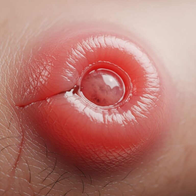
When demodicosis becomes symptomatic, the pathological changes result in characteristic clinical manifestations. The typical presentation includes follicular scales, erythema, and sensitive skin. Patients frequently develop pruritus (itching), which may range from mild to severe, and skin xerosis (dryness) as the mites and inflammatory mediators disrupt normal sebaceous gland function. Exfoliation and rash are common, with some patients experiencing postinflammatory hyperpigmentation resulting from mechanical damage and inflammatory processes that damage epidermal cells. The condition may manifest as macules, papules, eczema-like changes, or frank folliculitis, depending on the severity and extent of mite colonization.
A distinctive presentation described in the literature is demodex folliculorum or pityriasis folliculorum, characterized by a sandpaper-like texture of the skin due to follicular scaling and slight redness—a form of digitate keratosis that can cause significant irritation and burning sensations.
Ocular Demodicosis
The pathology of ocular demodicosis involves specific clinical manifestations related to meibomian gland dysfunction. Patients present with inflammation of the eyelid margin, characterized by lid thickening and follicular changes. The mite-induced pathology results in dysfunction of meibomian glands, leading to deficient lipid secretion essential for maintaining the lacrimal film. Complications include blepharitis (inflammation of the eyelid), chalazion (lipogranulomatous inflammation of meibomian glands), conjunctival inflammation, and decreased vision. Patients commonly report eye irritation, itching, and scaling of the eyelids, with some experiencing madarosis (loss of eyelashes) and eyestrain due to corneal irritation from abnormal tear composition.
Rosacea-Associated Demodicosis
Demodicosis frequently occurs in association with rosacea, representing a distinct pathological subtype. Rosacea-associated demodicosis presents as a drier form of rosacea, displaying superficial vesicles, pustules, and follicular scaling, in contrast to typical rosacea characterized by oily skin, absence of follicular scaling, and deeper tissue involvement. Studies have documented elevated Demodex prevalence in both erythemato-telangiectatic and papulopustular rosacea subtypes, with prevalence rates of 66.7% and 83.3% respectively. The cheek region is most heavily infested, followed by periocular areas, nose, chin, and mouth. Demodex may contribute to granuloma formation observed in papulopustular rosacea, creating a pathological cycle where the inflammatory environment promotes mite proliferation, which further drives inflammation.
Pathological Responses and Chronicity
A critical aspect of demodicosis pathology is its capacity to become chronic or recurrent. The condition can manifest as permanent lesions or recurring episodes of inflammation. This chronicity results from the self-perpetuating nature of the pathological process: mite proliferation triggers inflammation, which disrupts normal immune regulation and sebaceous gland function, creating an environment favorable for continued mite expansion. Without appropriate antidemodectic treatment targeting the underlying mite infestation, symptomatic management alone—including topical steroids, calcineurin inhibitors, systemic antibiotics, or antihistamines—frequently yields poor results and allows the pathological process to persist.
Diagnostic Pathology
Accurate diagnosis of demodicosis requires integration of clinical presentation with parasitological findings. The pathological diagnosis is established through several methods:
- Relevant clinical symptoms: The presence of characteristic signs including follicular scales, erythema, pruritus, and eyelid involvement.
- Mite density assessment: Demodicosis is defined pathologically as a mite concentration exceeding 5 per cm² or penetration of mites into the dermis. Standardized skin surface biopsy or direct microscopic examination identifies mite presence and quantifies density.
- Skin biopsy: Histopathological examination reveals mites within hair follicles, surrounding inflammatory infiltrates, and associated pathological changes including hyperkeratinization and epithelial hyperplasia.
- Therapeutic confirmation: Clinical resolution of signs and symptoms following acaricidal treatment provides pathological confirmation of the diagnosis.
Pathological Complications and Secondary Changes
Untreated or severe demodicosis can progress to significant pathological complications. Secondary bacterial infection frequently occurs due to follicular obstruction and compromised skin barrier function, potentially leading to deeper skin infections with abscess formation. Lymph node enlargement may occur in response to secondary infection. Postinflammatory changes, including hyperpigmentation and hypopigmentation, can persist long after resolution of active mite infestation due to damage to melanocyte function. The chronic inflammatory state can also exacerbate associated conditions such as rosacea or seborrheic dermatitis, creating complex pathological presentations requiring careful clinical assessment.
Pathophysiology of Pigmented Demodicosis
A rare but notable pathological variant is pigmented demodicosis, in which patients present with progressively pigmented plaques over facial areas, particularly the cheeks and eyelids. The pathological mechanism involves mechanical damage and local inflammatory processes triggered by mites in hair follicles, causing damage to epidermal cells and resulting in postinflammatory hyperpigmentation. This variant highlights the diversity of pathological presentations possible with demodicosis and underscores the importance of considering mite infestation in cases of facial pigmentation changes.
Management Implications of Pathology
Understanding the pathological mechanisms of demodicosis directly informs treatment strategy. Recognition that the condition results from excessive mite proliferation rather than infection per se explains why antimicrobial agents alone are ineffective. Effective management requires acaricidal therapy, with topical ivermectin representing the gold standard treatment, demonstrating marked improvement in pathological signs and symptoms within weeks to months. Maintenance therapy is often necessary due to the chronic nature of the pathology. Adjunctive measures targeting the predisposing environment include twice-daily facial cleansing, avoidance of greasy cosmetics and oil-based cleansers, and regular exfoliation to remove dead skin cells that serve as nutrients for mites. Caution is advised against excessive use of creams or moisturizers, as lipid-rich products provide additional nutrition for Demodex.
Frequently Asked Questions About Demodicosis Pathology
Q: Are Demodex mites normal on human skin?
A: Yes, Demodex mites are normal inhabitants of human skin in most individuals and typically cause no symptoms. Demodicosis develops only when mite populations proliferate excessively due to immunosuppression, skin disease, or other predisposing factors.
Q: What distinguishes primary from secondary demodicosis?
A: Primary demodicosis occurs in otherwise healthy individuals with mite overgrowth, while secondary demodicosis develops in patients with underlying inflammatory skin disorders or systemic diseases that promote abnormal mite proliferation.
Q: How is demodicosis definitively diagnosed?
A: Diagnosis requires clinical symptoms, demonstrated high mite density (>5 per cm²) through skin surface biopsy or microscopic examination, and clinical resolution following acaricidal treatment.
Q: Why do topical steroids alone fail to treat demodicosis?
A: Because demodicosis pathology fundamentally involves parasitic mite proliferation rather than simple inflammation, anti-inflammatory agents cannot address the underlying cause. Acaricidal therapy targeting the mites is essential for resolution.
Q: Is demodicosis contagious between humans?
A: Direct human-to-human transmission is not the primary route of spread. Rather, demodicosis develops when individual factors permit mite proliferation in naturally colonized skin.
Q: What is the role of sebaceous glands in demodicosis pathology?
A: Demodex mites feed on sebum and epidermal cells, concentrating in sebum-rich areas. Sebaceous gland dysfunction or hyperplasia promotes mite proliferation and is a recognized predisposing factor for demodicosis development.
References
- Primary facial demodicosis as a health problem and aesthetic consideration — National Center for Biotechnology Information. 2021-02-15. https://pmc.ncbi.nlm.nih.gov/articles/PMC7891371/
- Striking case of pigmented demodicosis — Skin Health and Disease, Oxford Academic. 2024-01-18. https://academic.oup.com/skinhd/article/5/5/386/8210514
- Beyond the Surface: Understanding Demodex and Its Link to Rosacea and Other Conditions — National Center for Biotechnology Information. 2024-06-10. https://pmc.ncbi.nlm.nih.gov/articles/PMC11213710/
- Demodex, Demodicosis: Clinical Features and Pathology — DermNet New Zealand. Accessed 2024. https://dermnetnz.org/topics/demodex
- Demodicosis — Classification, Treatment and Its Occurrence in Various Dermatological Conditions — Forum Dermatologicum, Via Medica. 2023-08-15. https://journals.viamedica.pl/forum_dermatologicum/article/view/65983
- Demodex (Face Mites): Folliculorum, Brevis & Treatment — Cleveland Clinic. 2024-01-20. https://my.clevelandclinic.org/health/diseases/22775-demodex-face-mites
Similar Articles


Read full bio of Sneha Tete