Dental Sinus: Definition, Causes, Symptoms & Treatment Guide
Understanding dental sinus: causes, diagnosis, and effective treatments for this common yet often misdiagnosed condition.

A
dental sinus
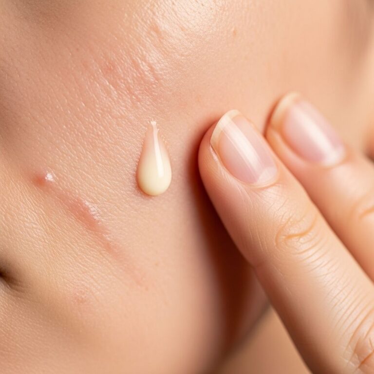
is a type of fistula characterised by a dimple, nodule, lump or pimple-like lesion with occasional discharge that connects to an infected tooth via a sinus tract through the bone and soft tissues of the face. It most commonly presents on the face just below the lower eyelid or above the upper canine teeth. Dental sinus tracts can occur anywhere on the face or neck depending on which tooth is infected. They may mimic more concerning pathologies such as basal cell carcinoma and therefore must be assessed by a dermatologist.What is the cause of a dental sinus?
Dental sinus tracts are caused by chronic dental infections (pulpitis or periodontitis) that have not been treated in a timely manner. The infection spreads from the root tip of the affected tooth along the path of least resistance through the bone and into the soft tissues to form an abscess. Over time, the pus-filled abscess spontaneously drains through the skin or oral mucosa to form a sinus tract. The most common causes are periapical abscesses, periodontal abscesses and dental implants.
- Periapical abscess: Necrotic dental pulp (pulpitis) from untreated dental caries or trauma leads to infection of the apical dental pulp and periodontium.
- Periodontal abscess: Infection of the gums and periodontal ligament from chronic periodontitis.
- Dental implants: Failed dental implants or peri-implantitis.
Upper teeth most commonly cause sinus tracts below the lower eyelid or above the medial aspect of the eyebrow. Maxillary canine and premolar teeth drain to the face above the upper lip or maxillary labial sulcus. Mandibular teeth drain to the submandibular space, causing swelling below the jawline.
Clinical features of dental sinus
Dental sinuses are usually asymptomatic but may be intermittently painful, swollen, red or itchy. They may discharge pus or serous fluid. The overlying skin is usually intact. There is often a visible dimple or nodule. The tract may contain a piece of inspissated dental debris (dentalis). There is no pain on palpation of the tooth unless acute infection supervenes. There are usually no signs of systemic infection.


Diagnosis of dental sinus
The diagnosis of dental sinus is confirmed by identifying the dental cause on dental radiographs. Orthopantomogram (OPG) or intraoral periapical radiographs (IOPA) demonstrate periapical radiolucency (dark area) or periodontal bone loss. Rarely, a gutta-percha point can be inserted into the sinus tract and its position relative to the apices of the teeth confirmed radiographically. Biopsy of the lesion is not necessary unless the clinical diagnosis is in doubt.
Differential diagnosis of dental sinus
Skin lesions on the face that resemble dental sinus include the following:
- Basal cell carcinoma: Nodule or ulcer with a pearly rolled edge, telangiectasia and lack of dental pathology.
- Squamous cell carcinoma: Scaly keratotic plaque or ulcer.
- Melanoma: Irregular pigmented lesion.
- Pyogenic granuloma: Bright red papule that rapidly enlarges.
- Actinic granuloma: Annular lesion on sun-exposed skin.
- Foreign body reaction: History of trauma or skin piercing.
Who should examine a dental sinus? A dentist should examine all dental sinuses to determine the dental cause and initiate dental treatment. A dermatologist should biopsy lesions that do not have an obvious dental cause.
What is the treatment for dental sinus?
Removal of the entire tooth (**extraction**) or necrotic dental pulp (**root canal / endodontic treatment**) is the only successful treatment for a dental sinus. Antibiotics such as penicillin or metronidazole may be also required. The sinus will usually heal 1–2 weeks after extraction or successful endodontic treatment. There may be residual scarring if biopsies or surgery had been performed. Otherwise there might be a slight dimple or skin surface colour change that usually improves with time.
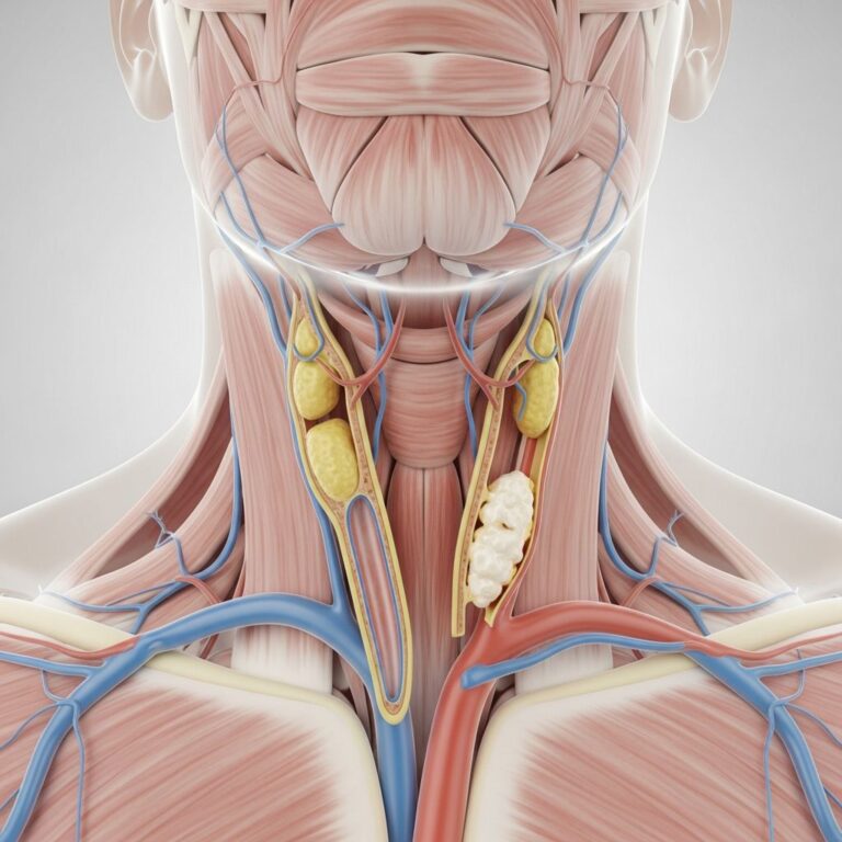
Odontogenic Rhinosinusitis (ORS)
Longstanding dental infections can spread directly into the maxillary sinus causing
odontogenic maxillary sinusitis
orodontogenic rhinosinusitis (ORS)
. This is the most common cause of unilateral maxillary sinusitis (40–45% of cases). Symptoms include nasal obstruction, purulent rhinorrhoea, post-nasal drip, facial pain/pressure, headache, and foul-smelling breath. Dental symptoms such as toothache may be absent. Diagnosis requires dental and ENT evaluation with CT imaging showing mucosal thickening, periapical lucency, and oroantral communication. Treatment combines dental intervention (extraction/root canal) with antibiotics, nasal steroids, saline irrigation, and sometimes functional endoscopic sinus surgery (FESS).| Feature | Rhinogenic Sinusitis | Odontogenic Sinusitis |
|---|---|---|
| Origin | Nasal/sinus mucosa | Dental infection |
| Prevalence | 55–60% | 40–45% |
| Symptoms | Bilateral, seasonal | Unilateral, persistent |
| Dental signs | Absent | Present (e.g., periapical lucency) |
| Imaging | Mucosal thickening | Bone erosion, tooth pathology |
Prevention of Dental Sinus
Regular dental check-ups, prompt treatment of caries and periodontitis, good oral hygiene, and avoiding trauma prevent dental infections that lead to sinuses. Patients with persistent facial lesions should seek dental evaluation early.
Frequently Asked Questions (FAQs)
Q: Can a dental sinus heal without treatment?
A: No, dental sinuses persist until the underlying dental infection is treated via extraction or root canal therapy.
Q: Is a dental sinus cancerous?
A: Rarely, but it must be differentiated from skin cancers like basal cell carcinoma via biopsy if no dental cause is found.
Q: How long does a dental sinus take to heal?
A: Typically 1–2 weeks after successful dental treatment, though scarring may remain.
Q: What antibiotics are used for dental sinus?
A: Penicillin or metronidazole, alongside definitive dental treatment.
Q: Can dental implants cause sinus tracts?
A: Yes, failed implants or peri-implantitis can lead to sinus formation.
Advanced Diagnostic Approaches
Cone-beam CT (CBCT) provides superior detail for detecting subtle periapical lesions compared to standard radiographs. Flexible nasal endoscopy visualises pus draining from the maxillary ostium in ORS cases. Bacterial cultures guide antibiotic choice, as odontogenic infections often involve anaerobes like Prevotella and Fusobacterium.
Treatment Outcomes and Complications
Early intervention yields >90% resolution rates. Untreated cases risk oroantral fistula, chronic ORS, or facial cellulitis. Multidisciplinary care (dentist + ENT + dermatologist) optimises outcomes. Endoscopic surgery (FESS) is preferred over Caldwell-Luc for refractory ORS, minimising morbidity.
Expanding on clinical manifestations, patients often report nasal obstruction as the initial symptom in ORS, progressing to purulent rhinorrhoea (66.7% prevalence) and hyposmia. Oral exam reveals vestibular oedema, tender maxillary wall percussion, and root fractures. Radiology confirms diagnosis: periapical osteitis appears as apical radiolucency; Schneiderian membrane perforation indicates direct spread.
Therapeutic algorithms prioritise dental source elimination: endodontic retreatment preserves teeth where possible, involving biomechanical cleaning, disinfection, and hermetic filling. Extraction is definitive for untreatable cases or root tips in the sinus. Adjunctive therapies include saline rinses, intranasal corticosteroids, and prolonged antibiotics (2–4 weeks) targeting polymicrobial flora.
In paediatric populations, trauma-induced pulp necrosis is common; adults more often present with caries-related disease. Immunocompromised patients risk rapid dissemination. Prognosis excels with prompt care; recurrence signals incomplete dental resolution.
Epidemiology underscores underdiagnosis: up to 45% of unilateral maxillary sinusitis is odontogenic, yet <20% initially recognised as such. Heightened awareness via interdisciplinary protocols reduces morbidity.
References
- Tooth-Related (Odontogenic) Sinusitis — Henry Ford Health. 2023. https://www.henryford.com/Services/Sinus/Conditions/Tooth-Related-Odontogenic-Sinusitis
- Odontogenic sinusitis: causes, diagnosis and treatment — Top Doctors. 2024. https://www.topdoctors.co.uk/medical-articles/odontogenic-sinusitis-causes-diagnosis-and-treatment/
- Odontogenic Sinusitis: How your teeth can cause sinusitis — Dr. Glicksman. 2023. https://www.doctorglicksman.com/post/odontogenic-sinusitis-how-your-teeth-can-cause-sinusitis
- Odontogenic Sinusitis: From Diagnosis to Treatment Possibilities — PMC (Peer-reviewed). 2022-07-20. https://pmc.ncbi.nlm.nih.gov/articles/PMC9319441/
- Sinusitis, dental infection or both — UT Health San Antonio. 2023. https://news.uthscsa.edu/sinusitis-dental-infection-or-both/
- Dental sinus — DermNet NZ. 2024. https://dermnetnz.org/topics/dental-sinus
Similar Articles


Read full bio of medha deb