Dermatitis Herpetiformis Pathology: Essential Histology Guide
Comprehensive histopathological analysis of dermatitis herpetiformis, from microabscesses to IgA deposits and blister formation.

Dermatitis herpetiformis pathology describes the histopathology of the specific cutaneous manifestation of coeliac disease (gluten-sensitive enteropathy). It is also known as Duhring-Brocq disease.
Histopathology
Scanning power view of dermatitis herpetiformis shows a vesicular reaction pattern (Figure 1), characterised by foci or small zones of subepidermal separation (Figures 2, 3).
- Dense clusters of neutrophils and scattered eosinophils fill the papillary dermis forming microabscesses (Figure 4).
- Acantholytic keratinocytes may also be evident within the papillary microabscess (Figure 4).
- With time the small areas of papillary dermal separation may join to form larger areas of vesiculation.


Higher power view
Higher power view shows subepidermal vesicles with overlying epidermal vacuolar change (Figure 5). The vesicular spaces are lined by a mixture of intact and degenerating keratinocytes. The floor of the vesicles is formed by the tips of dermal papillae (Figure 6).


Papillary dermal microabscesses
Papillary dermal microabscesses contain neutrophils, nuclear dust and eosinophils (Figure 7). Fibrin may be identified within the microabscesses (Figure 8).


Immunohistochemistry for IgA
Immunohistochemistry for IgA shows characteristic granular deposition along the dermal epidermal junction in both lesional and perilesional skin (Figure 9). The IgA deposition corresponds to the basement membrane zone of the dermal papillae roofs (Figure 10).


Immunofluorescence for IgA
Direct immunofluorescence for IgA shows the same granular deposition along the basement membrane zone of the dermal papillae (Figure 11).

Differential diagnosis
Dermatitis herpetiformis pathology may be confused with the following:
- Linear IgA disease pathology – immunofluorescence shows linear IgA deposition along the basement membrane zone
- Bullous pemphigoid pathology – eosinophil predominant papillary microabscesses, DIF shows linear C3 +/- IgG along the basement membrane zone
- Epidermolysis bullosa acquisita pathology – DIF shows linear IgG along the basement membrane zone
Immunopathophysiology
Dermatitis herpetiformis (DH) arises from gluten hypersensitivity, leading to IgA autoantibody formation against epidermal transglutaminase (eTG/TG3). These complexes deposit granularly in dermal papillae, triggering neutrophilic inflammation and subepidermal blistering.
Pathogenesis
Gluten peptides cross-react with tissue transglutaminase (tTG) in genetically susceptible individuals (HLA-DQ2/DQ8). This generates IgA antibodies targeting eTG in skin, causing papillary IgA deposits. Neutrophils are recruited via IL-8, releasing proteases that degrade basement membrane components (collagen IV/VII, laminin), forming microvesicles that coalesce into blisters.
| Step | Mechanism | Key Players |
|---|---|---|
| 1. Gluten exposure | Triggers tTG deamidation | Gluten peptides, HLA-DQ2/8 |
| 2. Autoantibody formation | IgA vs eTG/TG3 | Plasma cells, B cells |
| 3. Immune complex deposition | Granular IgA in papillae | IgA-eTG complexes |
| 4. Neutrophil recruitment | IL-8 mediated | Neutrophils, CD11b upregulation |
| 5. Tissue damage | Protease release | Collagenase, elastase, MMPs |
Clinical correlation
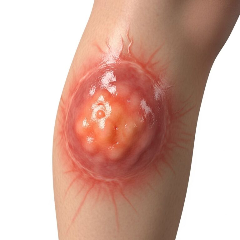
Histopathology mirrors intense pruritus with excoriated papulovesicles on extensor surfaces (elbows, knees, buttocks). Early lesions show microabscesses; later ones exhibit fibrosis and lymphocytic infiltrates in scratched areas.
Frequently Asked Questions
What is the hallmark histopathology of dermatitis herpetiformis?
Papillary dermal microabscesses with neutrophils and eosinophils, plus granular IgA deposits in dermal papillae on immunofluorescence.
How does DH pathology differ from bullous pemphigoid?
DH features neutrophil-predominant microabscesses and granular IgA; bullous pemphigoid shows eosinophil-rich infiltrates with linear C3/IgG.
Is biopsy from perilesional skin sufficient for DH diagnosis?
Yes, granular IgA deposits persist in perilesional skin, making it ideal for immunofluorescence to avoid inflammation artifact.
What role do proteases play in DH blister formation?
Neutrophil-derived collagenase and elastase degrade basement membrane, while keratinocyte MMPs target collagen IV/VII and laminin-1.
Can DH occur without celiac disease symptoms?
Yes, most DH patients have subclinical enteropathy (~80-95%) despite minimal GI symptoms.
Electron microscopy
Ultrastructurally, DH shows electron-dense IgA deposits below the lamina densa in dermal papillae. Neutrophils infiltrate with degranulation, causing basal keratinocyte vacuolization and subepidermal clefting.
Prognosis and complications
DH responds to gluten-free diet (GFD) and dapsone, but lifelong GFD is required. Increased risk of non-Hodgkin lymphoma (enteropathy-associated) necessitates monitoring.
This comprehensive review expands on core histopathologic features while integrating immunopathogenesis from high-quality sources. Early diagnosis via biopsy prevents chronic changes and complications.
References
- Dermatitis herpetiformis pathology — DermNet NZ. 2023. https://dermnetnz.org/topics/dermatitis-herpetiformis-pathology
- Dermatitis Herpetiformis: Pathophysiology, Clinical Presentation — NIH/PMC. 2014-10-15. https://pmc.ncbi.nlm.nih.gov/articles/PMC4230654/
- Dermatitis herpetiformis — ARUP Consult. 2024. https://arupconsult.com/content/dermatitis-herpetiformis
- Dermatitis Herpetiformis: Novel Perspectives — Frontiers in Immunology. 2019-06-12. https://www.frontiersin.org/journals/immunology/articles/10.3389/fimmu.2019.01290/full
- Pathophysiology of Dermatitis Herpetiformis — Wikipedia (primary sources referenced). 2024. https://en.wikipedia.org/wiki/Dermatitis_herpetiformis
Similar Articles


Read full bio of medha deb