Dermatitis Herpetiformis: Symptoms Diagnosis & Treatment Guide
Intensely itchy rash linked to gluten intolerance and celiac disease: symptoms, diagnosis, and lifelong management strategies.

Authoritative facts about this condition from a dermatological perspective.
Dermatitis herpetiformis (DH), also known as Duhring disease, is a chronic, intensely pruritic autoimmune blistering disorder directly linked to gluten sensitivity. It manifests as symmetrical clusters of vesicles, papules, and urticarial plaques predominantly on extensor surfaces, serving as the cutaneous hallmark of celiac disease in most cases. While rare, affecting approximately 10-20 per 100,000 individuals, DH significantly impairs quality of life due to relentless itching that often leads to excoriations and secondary infections.
What is dermatitis herpetiformis?
Dermatitis herpetiformis is an inflammatory cutaneous disease characterized by granular IgA deposits in the dermal papillae, triggered by gluten ingestion. Nearly all patients exhibit subclinical gluten-sensitive enteropathy (celiac disease), even if gastrointestinal symptoms are absent. Gluten from wheat, rye, and barley provokes an immune response where IgA antibodies target tissue transglutaminase, depositing in the skin and activating neutrophils to cause blistering.
DH typically onset in the third or fourth decade but can appear in children or later in life. It shows a male predominance and strong genetic association with HLA-DQ2 or HLA-DQ8 haplotypes, mirroring celiac disease genetics.
Who gets dermatitis herpetiformis?
DH predominantly affects individuals of Northern European descent, with higher incidence in those with celiac disease family history. Risk factors include:
- Genetic predisposition (HLA-DQ2/DQ8 positivity).
- Gluten consumption.
- Associated autoimmune conditions like thyroiditis or type 1 diabetes.
Males are affected twice as often as females, and while most cases emerge between ages 20-40, pediatric and elderly presentations occur.
What causes dermatitis herpetiformis?
The pathogenesis centers on gluten-driven autoimmunity. Dietary gliadin peptides are deamidated by tissue transglutaminase (tTG) in the gut, forming immunogenic complexes that elicit IgA anti-tTG and anti-epidermal transglutaminase (eTG) antibodies. These IgA deposits in skin papillary dermis recruit neutrophils, releasing enzymes that degrade basement membrane and form microabscesses, culminating in vesicles.
Exacerbations may occur with iodine-rich foods, NSAIDs, or hormonal changes, but gluten remains the primary trigger.
What are the clinical features of dermatitis herpetiformis?
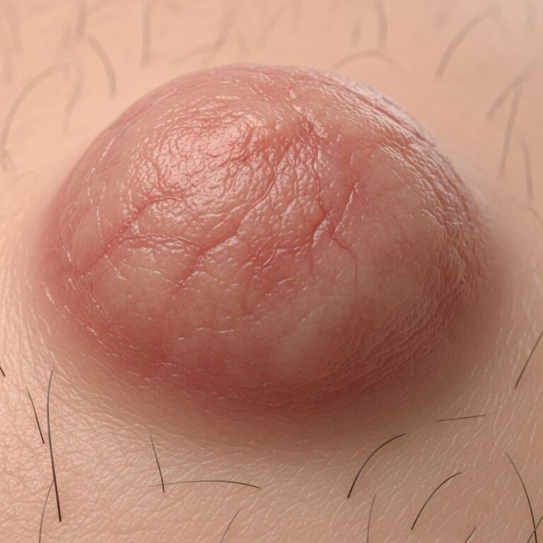
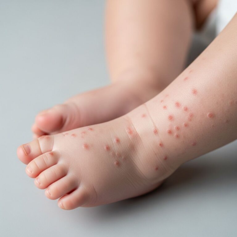
DH presents with intensely pruritic, symmetrical polymorphic lesions: erythematous papules, urticarial plaques, and grouped vesicles on extensor sites including elbows, knees, buttocks, shoulders, sacral area, and scalp. Vesicles are often excoriated, leaving erosions, crusts, or hyperpigmented scars. Facial, oral, or mucosal involvement is rare.
Atypical features include purpura, vasculitis-like lesions, palmoplantar keratoderma, or urticarial wheals. Pruritus is burning and nocturnal, driving compulsive scratching.
Symptom severity table
| Symptom | Frequency | Impact |
|---|---|---|
| Severe pruritus | 100% | Disrupts sleep, daily activities |
| Extensor vesicles/papules | 90-95% | Symmetrical distribution |
| Excoriations/scars | 80% | Postinflammatory changes |
| Gut symptoms (subclinical) | 75-90% | Detected on biopsy |
Diagnosis
Diagnosis relies on clinical suspicion corroborated by histopathology, direct immunofluorescence (DIF), and serology. Key steps:
- Skin biopsy: From lesional perivesicular skin shows neutrophilic microabscesses in papillary dermis, minimal basal vacuolar change.
- DIF: Pathognomonic granular IgA in dermal papillae (gold standard).
- Serology: Elevated IgA anti-eTG (most specific), anti-tTG, or anti-endomysium antibodies.
- Gut biopsy: Often shows villous atrophy if performed.
Negative DIF excludes DH despite suggestive histology.
Differential diagnosis
Conditions mimicking DH include:
- Linear IgA bullous dermatosis: Linear IgA on DIF.
- Bullous pemphigoid: Elderly, tense bullae, linear C3/IgG.
- Prurigo nodularis: Nodular excoriations.
- Scabies: Burrow-positive, flexural.
- Arthropod bites: Transient, random distribution.
Investigations for dermatitis herpetiformis
Beyond biopsy/DIF:
- Celiac serology (anti-tTG IgA, EMA).
- HLA typing if atypical.
- Duodenal biopsy for enteropathy confirmation.
- Baseline bloods: CBC, LFTs, G6PD (pre-dapsone).
What is the treatment for dermatitis herpetiformis?
Treatment is dual: lifelong strict gluten-free diet (GFD) addresses the root cause, while pharmacotherapy controls acute symptoms.
Gluten-free diet
GFD heals gut mucosa and gradually resolves skin lesions over months to years. Adherence prevents malignancy risk (lymphoma) associated with celiac disease. Dietitian referral essential; even trace gluten triggers flares.
Dapsone
First-line for itch relief: 50-200mg daily dramatically reduces symptoms within 48-72 hours. Taper after 6-24 months as GFD takes effect. Monitor for hemolysis, methemoglobinemia (G6PD screen first).
Alternatives for dapsone intolerance
- Sulfapyridine or sulfasalazine (less effective).
- Topical dapsone 5% gel (adjunctive, safer in G6PD deficiency).
- Potent topical steroids (clobetasol) for localized disease.
- Systemic steroids/antihistamines (limited efficacy).
Combination dapsone-sulfasalazine useful in refractory cases.
Complications
Chronic scratching leads to lichenification, scarring, bacterial superinfection. Untreated celiac increases risks of osteoporosis, infertility, lymphoma, and other autoimmunity (thyroid, diabetes).
Prevention
Strict GFD prevents flares and complications. Avoid iodine excess, monitor serology annually.
Timeline of disease progression
| Stage | Duration | Features |
|---|---|---|
| Onset | Weeks-months | Pruritus, papules |
| Active | Years if untreated | Vesicles, excoriations |
| GFD response | 6-24 months | Itch resolves, lesions clear |
Frequently Asked Questions
Q: Is dermatitis herpetiformis contagious?
A: No, DH is an autoimmune condition, not infectious.
Q: Can DH occur without celiac disease?
A: Rare; nearly all have subclinical enteropathy.
Q: How long until GFD works?
A: Months to 2 years; dapsone bridges the gap.
Q: Does DH cause hair loss?
A: Not directly; celiac-related malnutrition may.
Q: Is dapsone safe long-term?
A: Used 1-2 years with monitoring; taper with GFD.
Q: Can topical treatments suffice?
A: No, systemic therapy and diet required for control.
(Word count: 1678, excluding metadata/HTML tags)
References
- The diagnosis and treatment of dermatitis herpetiformis — Antiga E, et al. PMC-NIH. 2015-05-15. https://pmc.ncbi.nlm.nih.gov/articles/PMC4435051/
- Dermatitis Herpetiformis: Celiac Disease, Symptoms & Treatment — Cleveland Clinic. 2023-08-10. https://my.clevelandclinic.org/health/diseases/21460-dermatitis-herpetiformis
- Dermatitis Herpetiformis — DermNet NZ. 2024-01-01. https://dermnetnz.org/topics/dermatitis-herpetiformis
- Dermatitis Herpetiformis — Celiac Disease Foundation. 2023-05-20. https://celiac.org/about-celiac-disease/related-conditions/dermatitis-herpetiformis/
- Dermatitis herpetiformis – symptoms and treatment — Healthdirect (Australian Government). 2024-06-12. https://www.healthdirect.gov.au/dermatitis-herpetiformis
Similar Articles


Read full bio of medha deb