Dermatological Emergencies: 7 Conditions To Spot And Treat Fast
Essential guide to recognising and managing life-threatening skin conditions in clinical practice.

This continuing medical education module covers critical skin conditions that demand urgent recognition and management in clinical settings. Developed for health professionals, it emphasises rapid diagnosis and intervention to prevent morbidity and mortality.
Drug Eruptions
Drug eruptions represent a common yet potentially life-threatening dermatological emergency. Approximately 3% of hospitalised patients develop rashes attributable to adverse drug reactions. These eruptions often mimic infectious exanthems or underlying illnesses, complicating diagnosis. While most resolve upon drug withdrawal, severe forms like toxic epidermal necrolysis (TEN) and drug hypersensitivity syndrome (DHS) carry high mortality risks.
Clinical features vary widely, from morbilliform (measles-like) rashes to urticaria, fixed drug eruptions, and photosensitivity. Morbilliform eruptions, the most common type, present as symmetrical erythematous macules and papules starting on the trunk and spreading to extremities. They typically appear 7-14 days after drug initiation and improve within a week of discontinuation.
High-Risk Features
- Mucosal involvement
- Blistering or epidermal detachment
- Fever, lymphadenopathy, or organ dysfunction
- Progressive widespread erythema
- Eosinophilia or atypical lymphocytes on blood tests
Suspected culprits include antibiotics (amoxicillin, cephalosporins), anticonvulsants, NSAIDs, and allopurinol. A thorough drug history covering the past three months is essential, as reactions can occur even with long-term medications.
Types of Drug Eruptions
| Type | Features | Common Drugs |
|---|---|---|
| Exanthematous (Morbilliform) | Symmetrical trunk-to-limb rash, pruritic | Amoxicillin (>5%), Cephalexin, Trimethoprim |
| Urticarial | Wheals ± angioedema, may anaphylax | Penicillin, NSAIDs, Opioids |
| Fixed Drug Eruption | Oval plaques with central blister, recur in same site | Paracetamol, Fluconazole, Tetracyclines |
| Photosensitivity | Photodistributed erythema, sunburn-like | Doxycycline, Chlorpromazine, Quinine |
Management prioritises immediate withdrawal of the suspected drug and avoidance of cross-reacting agents. Symptomatic relief includes emollients, topical corticosteroids for mild cases, and oral antihistamines for urticaria. Severe reactions require hospitalisation, with systemic corticosteroids for DHS (e.g., prednisone 1-2 mg/kg/day). No reliable diagnostic tests exist; skin biopsy may show non-specific interface dermatitis or eosinophilic infiltrate.
Drug Hypersensitivity Syndrome (DHS)
DHS, also known as Drug Reaction with Eosinophilia and Systemic Symptoms (DRESS), manifests 2-8 weeks post-exposure with fever, rash, lymphadenopathy, and visceral involvement (liver, kidney, lung). Mortality approaches 10%. Aromatic anticonvulsants (carbamazepine, phenytoin) are classic triggers. Treatment involves drug cessation, systemic steroids, and supportive care.
Erythema Multiforme
Erythema multiforme (EM) is an acute, immune-mediated mucocutaneous disorder typically triggered by infections (Herpes simplex virus in 90% of cases) or drugs. It presents with ‘target’ lesions—concentric erythematous rings with central duskiness or blister—predominantly on acral sites. Classical EM involves skin without mucosal changes, distinguishing it from Stevens-Johnson Syndrome (SJS)/TEN spectrum.
Symptoms include pruritus or burning sensation. Diagnosis relies on clinical morphology; biopsy shows interface dermatitis with lymphocyte satellitosis. Management is supportive: wound care, analgesics, and antiviral therapy if HSV-associated. Most cases resolve in 2-4 weeks without scarring.
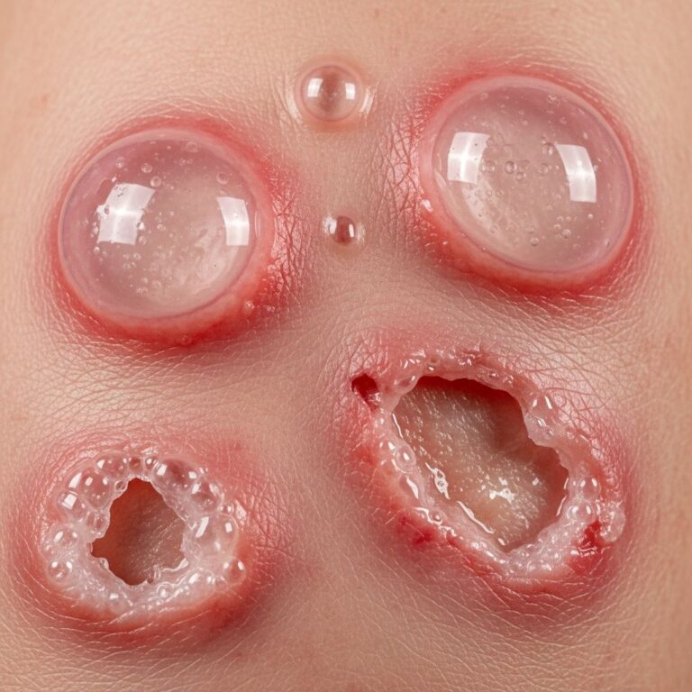
Toxic Epidermal Necrolysis (TEN)
TEN is a dermatological emergency with mortality up to 40%, characterised by widespread epidermal necrosis and detachment (>30% body surface area). It overlaps with SJS at 10-30% BSA involvement. Drugs account for 80% of cases, notably allopurinol, sulfonamides, and anticonvulsants, onset 1-3 weeks post-exposure.
Prodrome includes fever and flu-like symptoms, followed by painful erythema and sheet-like sloughing of epidermis. Positive Nikolsky sign (epidermis shears with gentle pressure) is pathognomonic. Urgent transfer to a burn unit is mandatory for barrier nursing, fluid resuscitation, and nutritional support. Controversial therapies include IVIG (1 g/kg/day x 3 days) and ciclosporin; corticosteroids are contraindicated due to infection risk.
Erythroderma
Erythroderma involves inflammatory erythema exceeding 90% BSA, with shedding of >800g/day scale. Causes include drugs (30%), pre-existing dermatoses (psoriasis, eczema), and malignancies (cutaneous T-cell lymphoma). Patients exhibit thermodysregulation, high-output cardiac failure risk, dehydration, and secondary infection.
Clinical triad: erythroderma, shivering, oedema. Labs show elevated IgE, anaemia, and eosinophilia. Management entails emollients, wet wraps, inpatient monitoring of fluids/electrolytes, and treating underlying cause (e.g., methotrexate for psoriasis). Mortality is 20-40% in elderly.
Neutrophilic Dermatoses
This group encompasses Sweet syndrome (acute febrile neutrophilic dermatosis), pyoderma gangrenosum, and others. Sweet syndrome features tender purple plaques/papules, fever, and neutrophilia, often post-infection or drug-induced (G-CSF). Pyoderma gangrenosum presents as painful ulcers with violaceous, undermined borders.
Diagnosis requires biopsy showing dense dermal neutrophilic infiltrate without vasculitis. Systemic corticosteroids (prednisone 1 mg/kg/day) are first-line; dapsone or biologics for refractory cases. Differentiation from infection is critical to avoid inappropriate antibiotics.
Urticaria and Angioedema
Urticaria (hives) and angioedema are mast cell-mediated, presenting as transient wheals and deep dermal swelling, respectively. Acute urticaria (<6 weeks) is often spontaneous or triggered by drugs/foods/infections. Chronic forms require investigation for inducible urticaria or autoimmunity.
Life-threatening if laryngeal involvement causes anaphylaxis. Second-generation H1-antihistamines (cetirizine 10mg BD) are mainstay; add H2-blockers or montelukast if partial response. Omalizumab or ciclosporin for refractory chronic urticaria. Epinephrine for anaphylaxis.
Blistering Diseases
Autoimmune blistering disorders like pemphigus vulgaris and bullous pemphigoid can present acutely. Pemphigus features flaccid blisters rupturing to painful erosions, acantholysis on biopsy. Bullous pemphigoid shows tense urticarial blisters in elderly. Rituximab and high-dose steroids are treatments; urgent dermatology referral essential.
Frequently Asked Questions (FAQs)
Q: How do I differentiate drug eruption from viral exanthem?
A: Drug eruptions often symmetrical, trunk-centric, with eosinophilia; viral exanthems more polymorphic, fever precedes rash.
Q: When to suspect TEN in a blistering patient?
A: Sheet-like epidermal detachment >10% BSA, mucous membrane involvement, recent drug exposure—transfer to ICU immediately.
Q: What is the first step in erythroderma management?
A: Admit for fluid/electrolyte monitoring, emollients, identify/treat underlying cause.
Q: Are skin tests reliable for drug allergies?
A: Rarely; history and drug withdrawal are key. Patch testing useful for some delayed reactions.
Q: How to manage anaphylaxis from urticaria?
A: IM epinephrine 0.3-0.5mg, airway support, antihistamines, corticosteroids.
References
- Drug eruptions – Dermatological emergencies — DermNet NZ. 2008 (updated). https://dermnetnz.org/cme/emergencies/drug-eruptions
- Dermatological emergencies – Contents page — DermNet NZ / University of Auckland. 2009. https://dermnetnz.org/cme/emergencies
- Toxic Epidermal Necrolysis: A Review — World Health Organization / Dermatology Guidelines. 2024-10-15. https://www.who.int/publications/i/item/9789240091475
- Guidelines for the Management of Stevens-Johnson Syndrome and Toxic Epidermal Necrolysis — British Association of Dermatologists. 2023-05-20. https://www.bad.org.uk/pils/sjs-ten/
- Drug Reaction with Eosinophilia and Systemic Symptoms (DRESS) — American Academy of Dermatology. 2025-01-10. https://www.aad.org/public/diseases/a-z/drug-reaction-eosinophilia
Similar Articles


Read full bio of medha deb