Dermatomes: 30 Essential Nerve Maps And Clinical Insights
Understanding dermatomes: segmental skin areas innervated by spinal nerves, essential for diagnosing neurological conditions.

A dermatome is defined as an area of skin primarily innervated by the dorsal root of a single spinal nerve, forming a segmental pattern that maps sensory distribution across the body. This concept, derived from Greek words ‘derma’ (skin) and ‘tome’ (segment), is fundamental in neurology and dermatology for pinpointing nerve root pathologies. Unlike the face, supplied by the trigeminal nerve (CN V), dermatomes cover the rest of the body via 31 pairs of spinal nerves (C2-C8, T1-T12, L1-L5, S1-S5), as C1 lacks a dorsal root in most individuals.
What are dermatomes?
Dermatomes represent the cutaneous sensory territories of spinal nerves, enabling clinicians to correlate skin sensations like pain, temperature, or touch loss with specific spinal levels. Each spinal nerve’s dorsal root ganglion contains sensory neuron cell bodies that relay signals from the skin to the spinal cord. Overlap between adjacent dermatomes (typically 2-3 nerves per skin point) ensures redundancy, preventing complete sensory loss from single nerve damage. This overlap complicates precise boundaries but aids functional resilience.
Embryologically, dermatomes arise from somites—segmental mesenchymal blocks flanking the neural tube. Each somite differentiates into sclerotome (vertebrae/ribs), myotome (muscles), and dermatome (skin dermis). During limb development, dermatomes elongate longitudinally due to limb rotation, contrasting the horizontal trunk bands. This results in ‘striped’ thoracic/abdominal patterns versus banded limb distributions.
Dermatome maps
Dermatome maps visualize these distributions, though variations exist due to individual anatomical differences and mapping methodologies (e.g., Foerster vs. Keegan-Garrett). No universal map is definitive; clinicians use them alongside patient exams for localization. Key maps include:
- Head/neck: Trigeminal nerve (V1-V3) for face; C2-C4 spinal nerves.
- Upper limbs: C5-T1, sequential from shoulder to fingers.
- Trunk: T2-T12, horizontal bands.
- Lower limbs/genitalia: L1-S3, longitudinal.
Head, face, and neck
The face follows trigeminal divisions: V1 (forehead), V2 (cheek), V3 (jaw). Neck dermatomes begin at C2 (occipital/supra-auricular), C3 (supraclavicular), C4 (acromioclavicular). These are tested at specific points, e.g., C2: 1 cm lateral to occipital protuberance.
Upper limbs
Innervated by C5-T1. C5 covers lateral shoulder/deltoid; C6, lateral forearm/thumb/index; C7, posterior forearm/middle fingers; C8, medial forearm/ring-little fingers; T1, medial arm/forearm. Maps differ: Keegan-Garrett shows continuous bands; Foerster, discontinuous at shoulder.
| Spinal Nerve | Upper Limb Region |
|---|---|
| C5 | Lateral arm, deltoid |
| C6 | Radial forearm, thumb, index finger |
| C7 | Posterior forearm, middle finger |
| C8 | Ulnar forearm, little finger |
| T1 | Medial forearm, distal arm |
Thorax and abdomen
T2-T12 form horizontal bands: T2 (axilla/upper chest), T4-T6 (pectoral/nipple), T10 (umbilicus), T12 (inguinal). Testing points: T6 at xiphisternum midclavicular line; T10 at umbilicus.
| Level | Location |
|---|---|
| T2 | Medial upper arm, pectoral |
| T4 | Nipple line |
| T6 | Xiphoid |
| T10 | Umbilicus |
| T12 | Inguinal ligament |
Lower limbs and genitalia
L1-L5, S1-S5: L1 (inguinal), L2-L3 (anterior thigh), L4 (medial leg), L5 (lateral leg/dorsum foot), S1 (posterior leg/lateral foot), S2-S4 (posterior thigh/perineum). Genitalia by S2-S4. Limb rotation creates longitudinal patterns.
| Spinal Nerve | Lower Limb Region |
|---|---|
| L1 | Upper thigh, groin |
| L2 | Anterior upper thigh |
| L3 | Anterior lower thigh, knee |
| L4 | Medial leg, medial foot |
| L5 | Lateral leg, dorsum foot, big toe |
| S1 | Posterior leg, lateral foot, little toe |
| S2-S4 | Posterior thigh, perineum |
Clinical significance
Dermatomes are pivotal for diagnosing spinal/nerve issues via sensory testing (light touch, pinprick, temperature). Patterns aid in:
- Radiculopathy: Pain/numbness in a single dermatome indicates root compression (e.g., C7 herniation: middle finger pain).
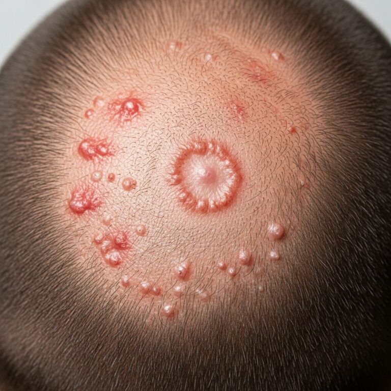
- Shingles (Herpes zoster): Varicella-zoster reactivation in dorsal root ganglia causes unilateral vesicular rash in 1-2 dermatomes (e.g., T5 thoracic).
- Spinal cord injury: Bilateral/multilevel loss below injury level.
- Neurogenic pain: Referred pain follows dermatomes.
Variations occur; maps are guides, not absolutes. Testing standardizes at midline points.
Frequently asked questions
What is a dermatome?
A dermatome is a skin area supplied by one spinal nerve’s dorsal root, mapping sensory innervation segmentally.
How many dermatomes are there?
Approximately 30: C2-8 (7), T1-12 (12), L1-5 (5), S1-5 (5), excluding C1 and face (trigeminal).
What causes shingles in a dermatome?
Varicella-zoster virus reactivation in a dorsal root ganglion, producing painful unilateral rash.
Can dermatomes overlap?
Yes, typically 2-3 nerves supply each skin point for redundancy.
How are dermatomes tested?
Using light touch, pinprick, or temperature on mapped points to detect sensory deficits.
Related topics
- Myotomes
- Spinal nerves
- Herpes zoster
- Radiculopathy
References
- Anatomy and dermatome map — Kenhub. 2023. https://www.kenhub.com/en/library/anatomy/dermatomes
- Dermatome (anatomy) — Wikipedia (informed by primary sources). 2024-01-15. https://en.wikipedia.org/wiki/Dermatome_(anatomy)
- Dermatomes — Physiopedia. 2024. https://www.physio-pedia.com/Dermatomes
- Anatomy, Skin, Dermatomes — StatPearls, NCBI Bookshelf (NIH). 2023-07-24. https://www.ncbi.nlm.nih.gov/books/NBK535401/
- Dermatomes: What They Are & Locations — Cleveland Clinic. 2023-11-01. https://my.clevelandclinic.org/health/body/24379-dermatomes
Similar Articles


Read full bio of medha deb