Diabetic Eye Disease: 3 Key Conditions And Prevention Tips
Understand risks, symptoms, detection, and treatments for diabetic eye disease to safeguard your vision effectively.

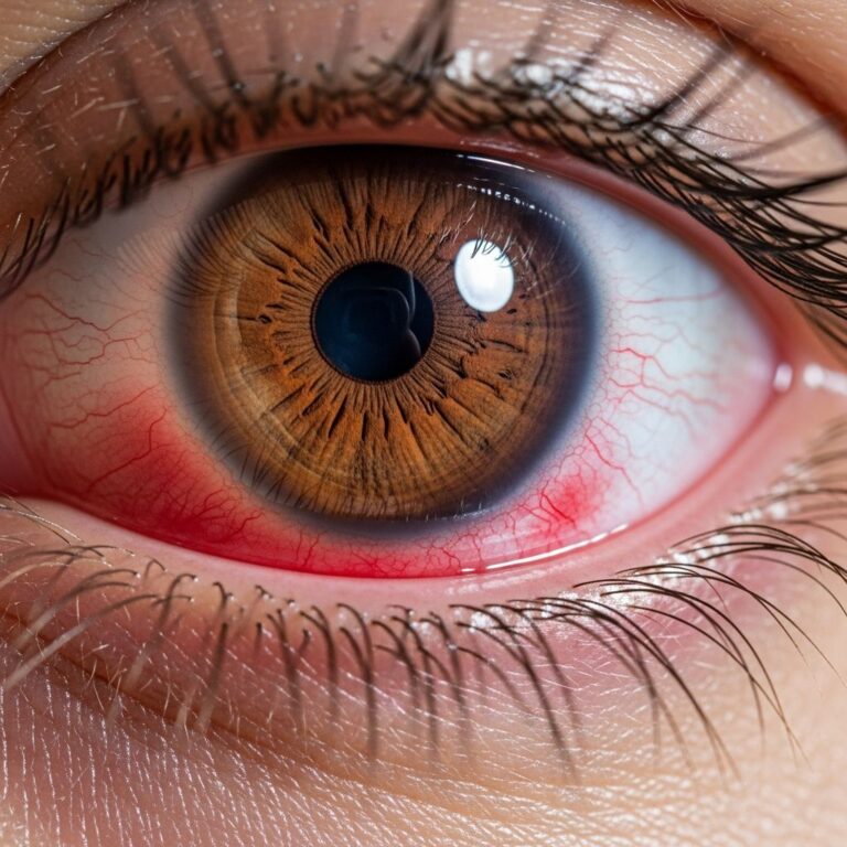
Diabetes significantly heightens the risk of vision impairment through conditions collectively termed diabetic eye disease. These issues stem from prolonged high blood sugar damaging delicate retinal blood vessels, potentially leading to blindness if unchecked. Early detection and diabetes control remain pivotal in averting severe outcomes.
The Link Between Diabetes and Vision Problems
High glucose levels over time weaken and narrow blood vessels supplying the retina, the eye’s light-sensitive layer. This triggers leakage, swelling, and abnormal vessel growth, disrupting clear vision. Approximately 30% of adults with diabetes develop some form of retinopathy, making it a leading cause of new blindness cases among working-age individuals.
Both type 1 and type 2 diabetes contribute, with duration of the disease being a key factor. Poor glycemic control accelerates damage, while factors like hypertension and high cholesterol compound risks. Managing these through lifestyle and medication forms the foundation of protection.
Core Types of Diabetic Eye Conditions
Diabetic eye disease manifests in distinct forms, each with unique characteristics and implications for sight.
- Non-Proliferative Diabetic Retinopathy (NPDR): The initial phase where vessels become leaky or blocked, causing microaneurysms, hemorrhages, and cotton wool spots. Vision may remain intact early on, but progression threatens central sight.
- Proliferative Diabetic Retinopathy (PDR): Advanced stage marked by new, fragile vessel growth (neovascularization) due to oxygen deprivation. These vessels bleed easily, leading to vitreous hemorrhage and potential retinal detachment.
- Diabetic Macular Edema (DME): Fluid buildup in the macula, the central retina responsible for sharp vision. It’s the primary reason for vision loss in diabetic patients, occurring independently or alongside retinopathy.
Other associated issues include accelerated cataracts and heightened glaucoma risk, further emphasizing comprehensive eye monitoring.
Recognizing Warning Signs Early
Many changes occur silently, underscoring the need for routine checks. When symptoms emerge, they include:
- Blurry or fluctuating central vision, signaling macular involvement.
- Sudden floaters or dark spots from bleeding.
- A shadowy curtain over vision, indicating retinal detachment.
- Difficulty reading or perceiving colors accurately.
These signs demand immediate professional evaluation to halt progression.
Essential Screening Protocols
Regular dilated eye examinations are non-negotiable for diabetes management. Guidelines vary by diabetes type:
| Diabetes Type | First Exam Timing | Follow-Up Frequency |
|---|---|---|
| Type 1 | Within 5 years of diagnosis | Annually |
| Type 2 | At diagnosis | Annually |
| Pregnancy with Diabetes | Pre-pregnancy or early first trimester | More frequent as needed |
Exams involve visual acuity tests, slit-lamp checks, intraocular pressure measurement, and dilated fundus inspection. Advanced tools like optical coherence tomography (OCT) provide cross-sectional retinal images to quantify swelling precisely.
Lifestyle and Medical Strategies for Prevention
Controlling blood sugar is paramount; each HbA1c percentage point drop reduces retinopathy risk substantially. Key steps include:
- Maintaining target HbA1c levels via diet, exercise, and medications.
- Controlling blood pressure and lipids to protect vessels.
- Avoiding smoking, which exacerbates vascular damage.
- Adopting a balanced diet rich in antioxidants for retinal health.
Consistent self-monitoring and collaboration with healthcare teams yield the best results.
Advanced Diagnostic Techniques
Beyond basic exams, specialists employ:
- Fundus Photography: Captures retinal images for progression tracking.
- Fluorescein Angiography: Dye injection highlights leaks and non-perfused areas.
- Ultrasonography: Assesses vitreous hemorrhage or detachment when views are obscured.
These enable tailored interventions before irreversible harm.
Treatment Options for Diabetic Retinopathy
Interventions target specific damage stages:
For NPDR and Early DME
Anti-VEGF injections such as aflibercept or ranibizumab block vessel growth factors, reducing leakage and edema. Administered monthly initially, they often restore vision acuity.
For Proliferative Stages
Panretinal photocoagulation (PRP) laser seals leaky vessels and curbs neovascularization, slashing severe vision loss risk by half, per landmark studies.
Surgical Interventions
Vitrectomy clears blood and scar tissue in vitreous hemorrhage or tractional detachment cases. Cataract removal enhances clarity when lens clouding coexists.
Treatment efficacy hinges on timely application; studies affirm observation for mild cases with good vision but prompt action for worsening.
Navigating Special Scenarios
Pregnant individuals with diabetes face rapid retinopathy worsening due to hormonal shifts. Frequent monitoring is advised. Similarly, those with kidney disease or post-laser history require adjusted schedules.
Role of Healthcare Collaboration
Optometrists and ophthalmologists play complementary roles: initial screenings by optometrists, referrals for suspected pathology. Primary care ensures systemic control.
Frequently Asked Questions
Can diabetic eye disease be reversed?
Early damage may stabilize or improve with control, but advanced scarring is permanent. Prevention trumps cure.
How often should I get eye exams if diabetic?
Annually minimum, or more if abnormalities present.
Does diet alone prevent retinopathy?
Diet aids control but pairs with medication and monitoring.
Are injections painful?
Topical anesthesia minimizes discomfort; side effects are rare.
What’s the success rate of laser treatment?
High-risk PDR sees 50% vision loss reduction over five years.
Empowering Long-Term Vision Health
Proactive steps transform diabetic eye disease from a looming threat to a manageable aspect of life. Commit to annual exams, blood sugar vigilance, and prompt symptom response for sustained sight.
References
- Diabetic Eye Disease Educators Guide — National Eye Institute (NEI). 2023-02-01. https://www.nei.nih.gov/sites/default/files/2023-02/Diabetic_Eye_Disease_Educators_Guide.pdf
- Promoting Eye Health — Centers for Disease Control and Prevention (CDC). Accessed 2026. https://www.cdc.gov/diabetes/hcp/clinical-guidance/promote-eye-health.html
- Diabetes-Related Retinopathy: Symptoms, Stages & Treatment — Cleveland Clinic. Accessed 2026. https://my.clevelandclinic.org/health/diseases/8591-diabetic-retinopathy
- Eye Health Resources — American Diabetes Association. Accessed 2026. https://professional.diabetes.org/clinical-support/eye-health-resources
- Diabetic Eye Disease: A Comprehensive Look at the Optometrist’s Role — Review of Optometry. Accessed 2026. https://www.reviewofoptometry.com/article/diabetic-eye-disease-a-comprehensive-look-at-the-optometrists-role
Similar Articles


Read full bio of medha deb